B ultrasonic equipment for detecting cholangiocarcinoma in pancreaticobiliary department

A technology for cholangiocarcinoma and B-ultrasound, applied in the fields of application, medical science, ultrasound/sonic wave/infrasonic wave diagnosis, etc., can solve the problems of low work and operation efficiency, low degree of automation, cross-infection of patients, etc., to prolong the service life, The effect of reducing safety hazards and improving measurement accuracy

Pending Publication Date: 2022-04-26
NINGBO FIRST HOSPITAL
View PDF0 Cites 0 Cited by
  • Summary
  • Abstract
  • Description
  • Claims
  • Application Information

AI Technical Summary

Problems solved by technology

[0002] B-ultrasound detector is a conventional medical equipment for cholangiocarcinoma detection. It generates incident ultrasonic waves (transmission waves) and receives reflected ultrasonic waves (echo waves) through B-ultrasound probes to detect the location and characteristics of tissues and organs. In the existing technology , when the B-ultrasound equipment uses the probe to check the patient, the attending doctor generally scans the parts of the human body that need to be checked manually. The image displayed on the display screen is judged, which affects the operation, resulting in low work and operation efficiency, and a low degree of automation. In the existing equipment, B-ultrasound equipment needs to apply coupling agent when using the probe, which is manually performed by the attending doctor. Smear, and the probe needs to be in contact with the patient's body. The attending doctor needs to test multiple patients every day, which may lead to the risk of doctors being infected with infectious diseases, and may cause cross-infection between patients

Method used


Image

  • B ultrasonic equipment for detecting cholangiocarcinoma in pancreaticobiliary department
  • B ultrasonic equipment for detecting cholangiocarcinoma in pancreaticobiliary department
  • B ultrasonic equipment for detecting cholangiocarcinoma in pancreaticobiliary department

Examples


Embodiment Construction

[0049] In order to further understand the features, technical means, and specific objectives and functions achieved by the present invention, the present invention will be further described in detail below in conjunction with the accompanying drawings and specific embodiments.

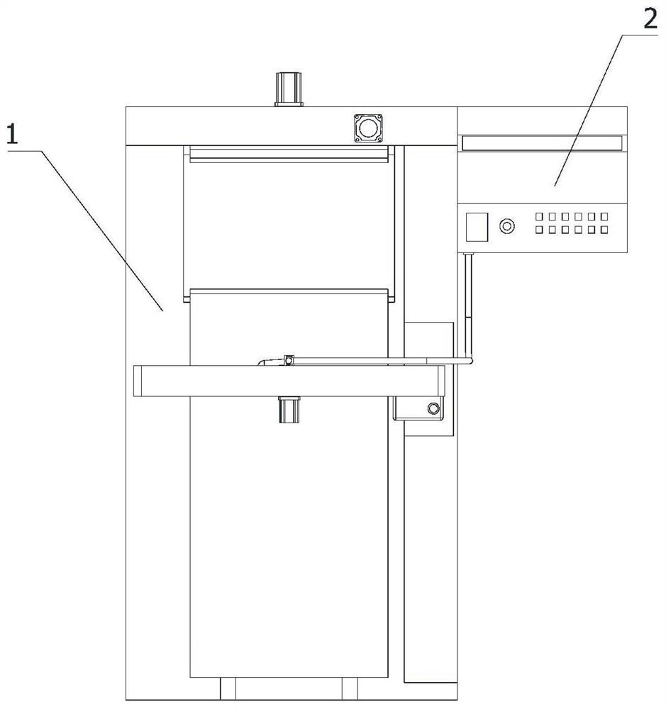
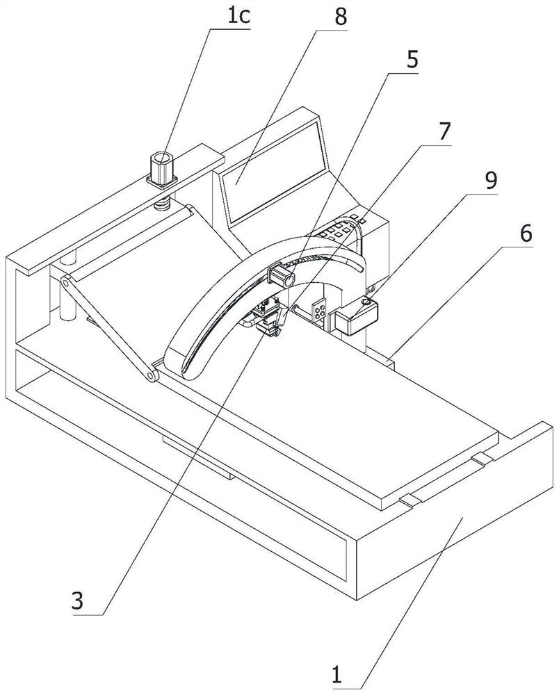
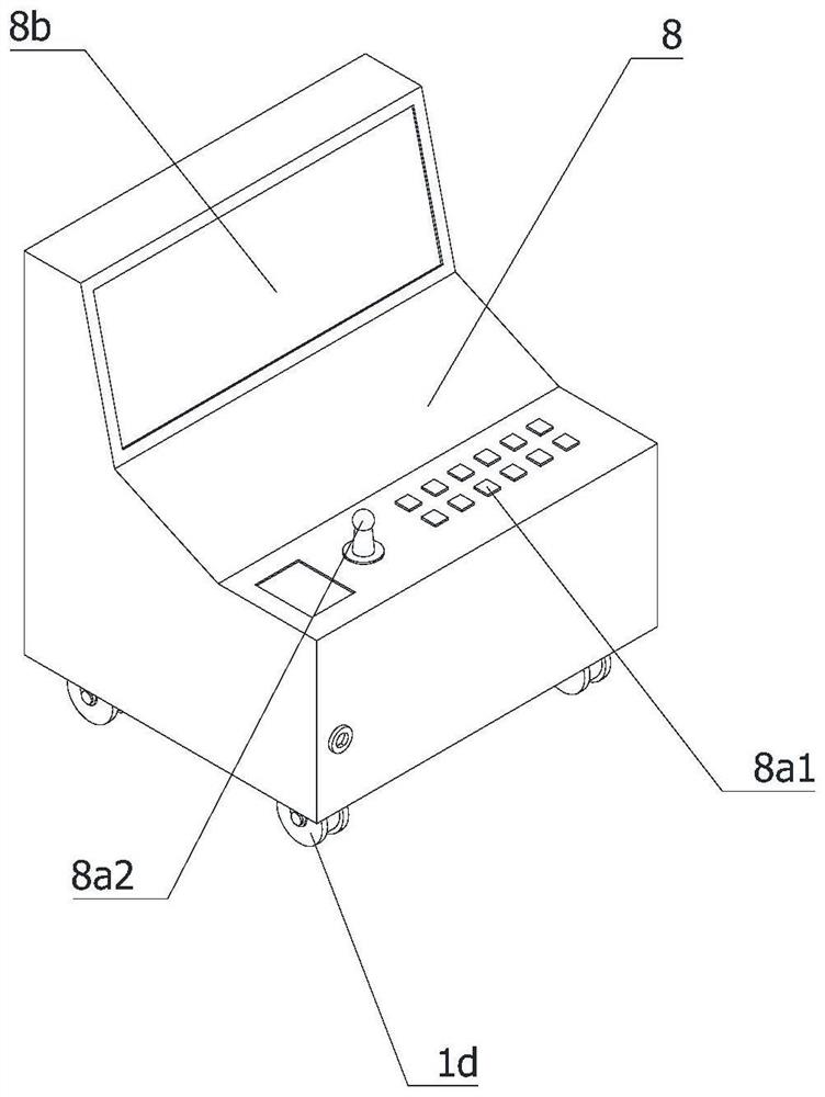
[0050] like Figure 1-15 As shown, this application provides:

[0051] A B-ultrasound device for detecting pancreaticobiliary cholangiocarcinoma, comprising a support base 1 and an ultrasonic mechanism 2, the ultrasonic mechanism 2 is located on the side of the support base 1, and the bottom of the support base 1 is provided with a The first slide rail 1a, the ultrasonic mechanism 2 includes a probe 3, an arc slide rail 4, a drive assembly 5 for the probe 3 to move along the arc slide rail 4, and a drive assembly 5 for driving the arc slide rail 4 along the first support seat 1 A slide rail 1a moves a translation assembly 6, a coupling agent application assembly 7, a control assembly 8, and a cleaning...


PUM


Abstract

The invention relates to the technical field of medical instruments and equipment, in particular to B-ultrasonic equipment for bile duct cancer detection in the pancreaticobiliary department. The B-ultrasonic equipment comprises an ultrasonic mechanism and a supporting seat, and a first sliding rail arranged in the length direction of the supporting seat is arranged at the bottom of the supporting seat; the ultrasonic mechanism comprises a probe, an arc-shaped sliding rail, a driving assembly, a translation assembly, a coupling agent smearing assembly, a control assembly and a cleaning assembly, through cooperation of the driving assembly, the translation assembly and the probe, a doctor can concentrate on observing the disease condition without distracting to control the probe, and the working efficiency is improved; the probe conforms to the radian of a human body when moving, the detection precision is improved, the comfort degree of a patient is improved when the patient is detected, a coupling agent is smeared on the moving path of the probe through the coupling agent smearing assembly, the probe is isolated from air, and the detection precision is improved; and the risk that patients and doctors are infected with infectious diseases due to the fact that the probe is not cleaned is avoided.

Description

technical field [0001] The invention relates to the technical field of medical devices, in particular to a B-ultrasound device for detecting pancreaticobiliary cholangiocarcinoma. Background technique [0002] B-ultrasound detector is a conventional medical equipment for cholangiocarcinoma detection. It generates incident ultrasonic waves (transmission waves) and receives reflected ultrasonic waves (echo waves) through B-ultrasound probes to detect the location and characteristics of tissues and organs. In the existing technology , when the B-ultrasound equipment uses the probe to check the patient, the attending doctor generally scans the parts of the human body that need to be checked manually. The image displayed on the display screen is judged, which affects the operation, resulting in low work and operation efficiency, and a low degree of automation. In the existing equipment, B-ultrasound equipment needs to apply coupling agent when using the probe, which is manually p...

Claims


Application Information

Patent Timeline
IPC IPC(8): A61B8/00
CPCA61B8/4209A61B8/4281A61B8/4422A61B8/40
Inventor 杨亮汪洁姜建帅
Owner NINGBO FIRST HOSPITAL
Why Eureka
Try Eureka