Vagina-implanted developing marking device for treating endometrial cancer

An endometrial cancer and marking device technology, which is applied in the field of imaging and marking devices for endometrial cancer treatment, can solve the problems of small uterine stump and inability to achieve accurate positioning of body surface line drawing, achieves favorable effects and is suitable for large-scale The effect of promoting use and reducing costs

Pending Publication Date: 2022-05-13
AFFILIATED CANCER HOSPITAL OF SHANDONG FIRST MEDICAL UNIV SHANDONG CANCER INST (SHANDONG CANCER HOSPITAL)
View PDF0 Cites 0 Cited by
  • Summary
  • Abstract
  • Description
  • Claims
  • Application Information

AI Technical Summary

Problems solved by technology

The uterine stump is small and deep, and it is impossible to accurately locate the body surface drawing line. Therefore,

Method used


Image

  • Vagina-implanted developing marking device for treating endometrial cancer
  • Vagina-implanted developing marking device for treating endometrial cancer
  • Vagina-implanted developing marking device for treating endometrial cancer

Examples


Embodiment 1

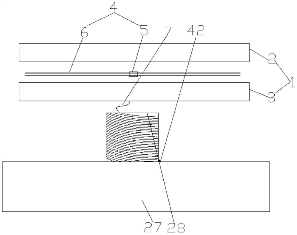
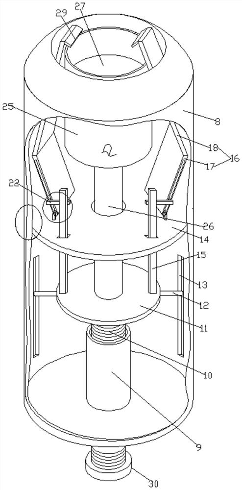
[0034] see Figure 1-7, according to an embodiment of the present invention, a transvaginally inserted endometrial cancer treatment development marking device includes a push mechanism and a development mechanism, the development mechanism includes a development cloth 1, and the development cloth 1 consists of an upper gauze piece 2 , the lower gauze piece 3 and the development strip 4, the development strip 4 is located between the upper gauze piece 2 and the lower gauze piece 3, the development strip 4 is composed of medical metal sheet 5 and development wire 6, The bottom end of the developing cloth 1 is fixed with a winding wire 7, the pushing mechanism includes a pipe 8, the top of the pipe 8 is open, the bottom of the pipe 8 is solid, and the middle part of the inner bottom of the pipe 8 is fixed. There is a sleeve 9, the inner wall of the sleeve 9 is in the shape of an internal thread, the sleeve 9 is threaded with a screw 10, the top of the screw 10 is connected with a...

Embodiment 2

[0037] see figure 1 , there are at least two developing filaments 6, and the developing filaments 6 are barium wire developing filaments.

Embodiment 3

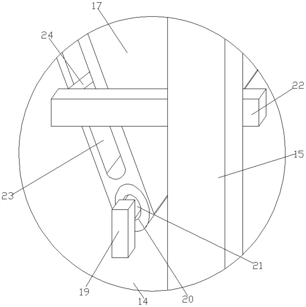
[0039] see Figure 4 , the bottom end of the screw 10 is fixed with a manual turntable 30, and the rotating mechanism includes a circular table 31 fixed at the bottom middle of the lift table 11, and the bottom middle of the circular table 31 is concavely provided with a T-shaped Turning slot 32 , the top end of the screw 10 is fixed with a connecting block 33 inside the T-shaped turning slot 32 .

[0040] Through the above solution of the present invention, the beneficial effect is that the top of the screw rod 10 is provided with a connecting block 33 inside the T-shaped turning groove 32, which drives the lifting platform 11 to go up and down without driving the lifting platform 11 to rotate.


PUM


Abstract

The invention discloses a transvaginal endometrial cancer treatment developing marking device which comprises a pushing mechanism and a developing mechanism, the developing mechanism comprises developing cloth, the developing cloth is composed of an upper gauze piece, a lower gauze piece and a developing strip, the developing strip is located between the upper gauze piece and the lower gauze piece, and the upper gauze piece and the lower gauze piece are arranged in parallel. The developing strip is formed by weaving medical metal sheets and developing wires, a winding wire is fixedly arranged at the bottom end of the developing cloth, the pushing mechanism comprises a pipeline, the top end of the pipeline is open, the bottom of the pipeline is solid, a sleeve is fixedly arranged in the middle of the inner bottom end of the pipeline, and the inner wall of the sleeve is in an internal thread shape. The sleeve is sleeved with a screw in a threaded mode, and the top end of the screw is connected with a lifting table. The positioning developing cloth has the beneficial effects that the developing cloth is left at the uterine stump through traction of the winding wire, the designed positioning developing cloth is put into the uterine stump through the vagina, and after work is finished, the developing cloth put into the uterine stump through the vagina can be pulled out through the designed winding wire.

Description

technical field [0001] The invention relates to the medical field, in particular to a developing marking device inserted through the vagina for treatment of endometrial cancer. Background technique [0002] Radiation therapy is performed after hysterectomy. The resection stump is located deep in the abdomen and overlaps with other tissues and organs. Radiation therapy cannot be directly positioned by the naked eye. Radiation therapy for other parts is usually positioned by drawing lines on the surface of the body. The uterine stump is small and deep, which makes it impossible to accurately locate the body surface. Therefore, this application proposes a vaginal-inserted imaging marking device for the treatment of endometrial cancer. [0003] Aiming at the problems in the related technologies, no effective solution has been proposed yet. Contents of the invention [0004] The object of the present invention is to provide a transvaginally implanted imaging marking device for...

Claims


Application Information

Patent Timeline
IPC IPC(8): A61N5/10
CPCA61N5/1016A61N5/1049A61N2005/1051
Inventor 谢鹏李晓琳刘乃富郭秋芬陈金龙王倩彭琛
Owner AFFILIATED CANCER HOSPITAL OF SHANDONG FIRST MEDICAL UNIV SHANDONG CANCER INST (SHANDONG CANCER HOSPITAL)
Why Eureka
Try Eureka