MR diagnostic device for imaging department and use method

A diagnostic device and imaging technology, applied in the field of nuclear magnetic diagnosis, can solve the problems of lack of classification diagnosis, difficult image processing, limitations of comprehensive diagnosis of patients' diseases, etc., to achieve the effect of improving accuracy

Inactive Publication Date: 2022-05-27
QINGDAO CENT HOSPITAL
View PDF0 Cites 0 Cited by
  • Summary
  • Abstract
  • Description
  • Claims
  • Application Information

AI Technical Summary

Problems solved by technology

[0005] Aiming at the above-mentioned shortcomings in the prior art, the present invention provides an MR diagnostic device and method for use in the imaging department, which solves the problem that when the imaging department recognizes the results of the patient's MRI images, most of the diagnostic work is performed by the service terminal based on the database. , there is no classified diagnosis and difficult images are difficult to deal with, so there are obvious limitations in the comprehensive diagnosis of patients' diseases

Method used


Image

  • MR diagnostic device for imaging department and use method
  • MR diagnostic device for imaging department and use method

Examples


Embodiment 1

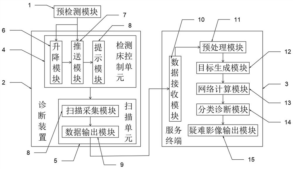
[0037] A method for using an MR diagnostic device for imaging department in this embodiment, such as figure 1 As shown, it includes a pre-detection module 1 and a diagnostic device 2, including:

[0038] The test bed control unit 4 composed of the lifting module 6 , the pushing module 7 and the prompting module 8 is used to push the tester into the diagnostic device 2 , and align the tester's to-be-tested area with the imaging area of ​​the diagnostic device 2 . MRI scan;

[0039] The scanning unit 5 composed of the scanning acquisition module 8 and the data output module 9 is used to scan the examiner on the detection bed, and output the sagittal image of the imaging area of ​​the examiner to the service terminal 3 for diagnosis processing through the data output module 9 ;

[0040] The service terminal 3 is used for receiving the examiner's MRI image data input by the diagnosis device 2 through the data receiving module 10, and processing and diagnosing the MRI image to de...

Embodiment 2

[0044] like figure 1 As shown, the pre-detection module 1 is used for pre-detecting whether the physical condition of the person to be detected meets the requirements of nuclear magnetic resonance detection, and whether there are metal objects and magnet objects on the person to be detected.

[0045] In this embodiment, the lifting module 6 is used to adjust the height of the test bed. When ready, the patient on the test bed is pushed to the NMR instrument through the push module 7, and the area to be tested of the tester is aligned with the imaging area of ​​the NMR instrument. The prompting module 8 is used for the operator to prompt the inspector information by voice, notify the inspector to perform ventilation or ventilating work during the scanning process, and automatically emit an alarm sound to prompt the inspector to complete the scanning operation.

[0046] The positions of the S-pole magnets and the N-pole magnets in the scanning unit 5 in this embodiment correspond...

Embodiment 3

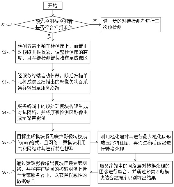
[0058] A method for using an MR diagnostic device in an imaging department, such as figure 2 As shown, the method is an implementation method of any one of the MR diagnostic devices for imaging departments in Embodiment 1 and Embodiment 2, and includes the following steps:

[0059] S1: Pre-detect whether the person to be tested meets the MRI scanning conditions, and if the person to be tested does not meet the MRI scanning conditions, further pre-test the person to be tested for a second time;

[0060] S2: According to the judgment in S1, if the subject to be tested meets the MRI scanning conditions, the subject needs to lie flat on the testing bed with his face facing the MRI apparatus, adjust the height of the testing bed, and push the part to be tested to the imaging area;

[0061] S3: Start the instrument through the service terminal, and then the scanning unit collects the image sagittal plane scanned in the imaging area and outputs it to the service terminal;

[0062] ...


PUM


Abstract

The invention relates to the technical field of nuclear magnetic diagnosis, in particular to an MR diagnosis device for the imaging department and a using method, the MR diagnosis device comprises a pre-detection module and a diagnosis device, and the MR diagnosis device comprises a detection bed control unit composed of a lifting module, a pushing module and a prompting module and used for pushing a detector into the diagnosis device, the scanning unit is composed of a scanning acquisition module and a data output module and is used for scanning the detector on the detection bed and outputting a sagittal plane image of the imaging area of the detector to the service terminal through the data output module for diagnosis processing; the generative adversarial network is constructed through the preprocessing module in the service terminal, so that the purpose of removing noise is achieved, subsequent disease classification diagnosis work and line meeting between difficult images and experts are facilitated, disease auxiliary diagnosis can be better achieved, and therefore the accuracy of nuclear magnetic detection is improved.

Description

technical field [0001] The invention relates to the technical field of nuclear magnetic diagnosis, in particular to an MR diagnosis device for imaging department and a method for using the same. Background technique [0002] Magnetic resonance imaging is a new medical imaging technology using the principle of nuclear magnetic resonance. diagnostic function. Compared with other auxiliary examination methods, MRI has the advantages of many imaging parameters, fast scanning speed, high tissue resolution, and clearer images. It can help doctors "see" the early lesions that are not easy to detect. A powerful tool for early screening of vascular diseases. [0003] However, in the current imaging department, when identifying the MRI results of patients, most of the service terminals carry out the diagnosis work based on the database, which does not have the classification diagnosis and the difficult images are difficult to deal with, so there are obvious limitations in the compre...

Claims


Application Information

Patent Timeline
Patent Type & Authority Applications(China)
IPC IPC(8): A61B5/055
CPCA61B5/055A61B5/0033
Inventor 刘桂芳郭成李常虹
Owner QINGDAO CENT HOSPITAL
Why Eureka
Try Eureka