Devices, systems, and methods for performing electroretinograms

A technology of electroretinogram and wearable equipment, applied in the direction of eye testing equipment, application, medical science, etc.

Pending Publication Date: 2022-08-05
THE GOVERNMENT OF THE UNITED STATES OF AMERICA AS REPRESENTED BY THE DEPT OF VETERANS AFFAIRS +1
View PDF0 Cites 0 Cited by
  • Summary
  • Abstract
  • Description
  • Claims
  • Application Information

AI Technical Summary

Problems solved by technology

This requires highly trained professionals to spend valuable time analyzing in busy clinics

Method used


Image

  • Devices, systems, and methods for performing electroretinograms
  • Devices, systems, and methods for performing electroretinograms
  • Devices, systems, and methods for performing electroretinograms

Examples


Embodiment Construction

[0040] The present invention may be understood more readily by reference to the following detailed description, examples, drawings, and claims, as well as the descriptions preceding and following them. However, unless otherwise indicated, before the present apparatus, system and / or method is disclosed and described, it is to be understood that this invention is not limited to the particular apparatus, system and / or method disclosed, as such may, of course, vary. It is also to be understood that the terminology used herein is for the purpose of describing particular aspects only and is not intended to be limiting.

[0041] The following description of the invention is provided as an effective teaching of the invention in its best, currently known embodiment. To that end, those skilled in the relevant art will recognize and appreciate that various changes can be made in the various aspects of the invention described herein, while still obtaining the beneficial results of the inv...


PUM


Abstract

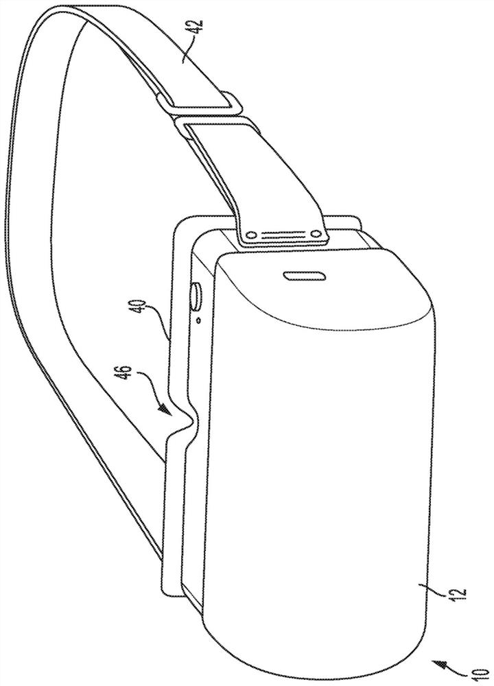
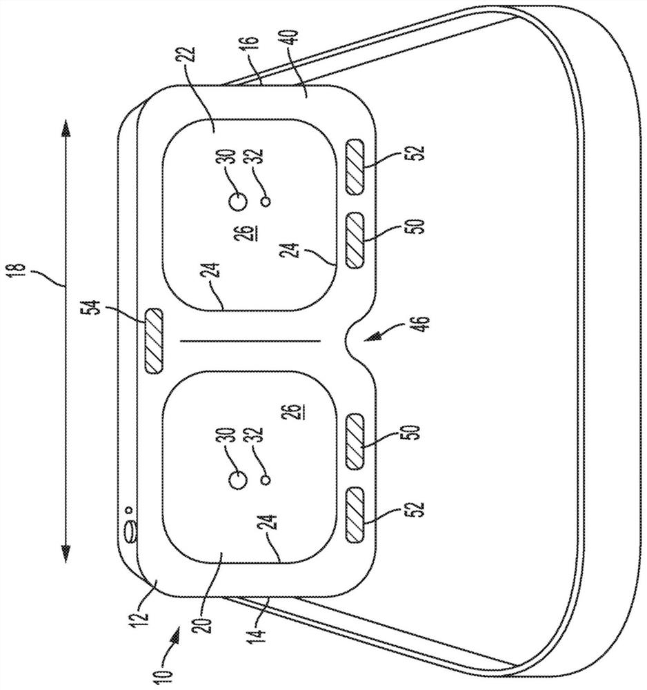
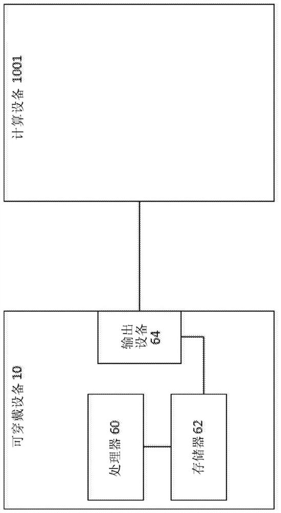
A wearable device for performing an electroretinogram examination on a wearer may have a housing defining a first compartment and a second compartment. Each of the first compartment and the second compartment may include: a stimulation light source; a focused light source; an active electrode configured to engage the skin of the wearer; and a reference electrode spaced apart from the active electrode and configured to engage the skin of the wearer. A processor may be communicatively coupled to the stimulation light source, the active electrode, and the reference electrode of each of the first compartment and the second compartment of the housing. A memory may be in communication with the processor. The apparatus may perform a method comprising: flashing the stimulation light source of the first compartment; and storing a signal from the active electrode of the first compartment. The housing may further include a ground electrode.

Description

[0001] CROSS-REFERENCE TO RELATED APPLICATIONS [0002] This application claims priority to and the benefit of US Provisional Patent Application No. 62 / 912,920, filed on October 9, 2019, the entire contents of which are incorporated herein by reference. technical field [0003] The present disclosure generally relates to systems, devices, and methods for testing retinal diseases. Background technique [0004] Diabetic retinopathy (DR) is the leading cause of blindness in working-age adults. Fundus photography is a routine method for diagnosing DR. However, by the time DR is evident on fundus photographs, irreversible damage and vision loss have already occurred. Electroretinography (ERG) is a powerful tool for recording retinal function, which is used in eye clinics to diagnose or monitor retinal diseases. By detecting defects in the rod drive pathway, ERG can screen for DR at an early stage, before vision-threatening damage occurs. Because: (a) the patient's eye requir...

Claims


Application Information

Patent Timeline
Patent Type & Authority Applications(China)
IPC IPC(8): A61B3/10A61B3/15
CPCA61B3/12A61B5/6831A61B5/256A61B5/297A61B5/6803A61B5/7275
Inventor K·切斯勒C·莫茨M·T·帕杜
Owner THE GOVERNMENT OF THE UNITED STATES OF AMERICA AS REPRESENTED BY THE DEPT OF VETERANS AFFAIRS
Why Eureka
Try Eureka