
Uterus shrinkage laplacian relief map instrument

A technology of uterine contraction Laplace and topography, which is applied in the field of medical instruments, can solve problems such as difficult to show the contraction phase of uterine muscle contraction direction, and the frequency band is not suitable for uterine myoelectricity, so as to ensure natural delivery and guarantee Maternal and infant safety and the effect of avoiding dystocia

Inactive Publication Date: 2006-11-08
TIANJIN UNIV
View PDF0 Cites 0 Cited by
  • Summary
  • Abstract
  • Description
  • Claims
  • Application Information

AI Technical Summary

Problems solved by technology

Although some people use ordinary electromyography machines to study uterine myoelectricity, the frequency band and gain of ordinary electromyography machines are not suitable for the measurement of uterine myoelectricity, and the display and recording methods of ordinary electromyography machines are one-dimensional. In the form of waveform, it is difficult to express the direction of uterine muscle contraction and the phase of contraction of each part

Method used


Image

  • Uterus shrinkage laplacian relief map instrument
  • Uterus shrinkage laplacian relief map instrument
  • Uterus shrinkage laplacian relief map instrument

Examples


Embodiment 1

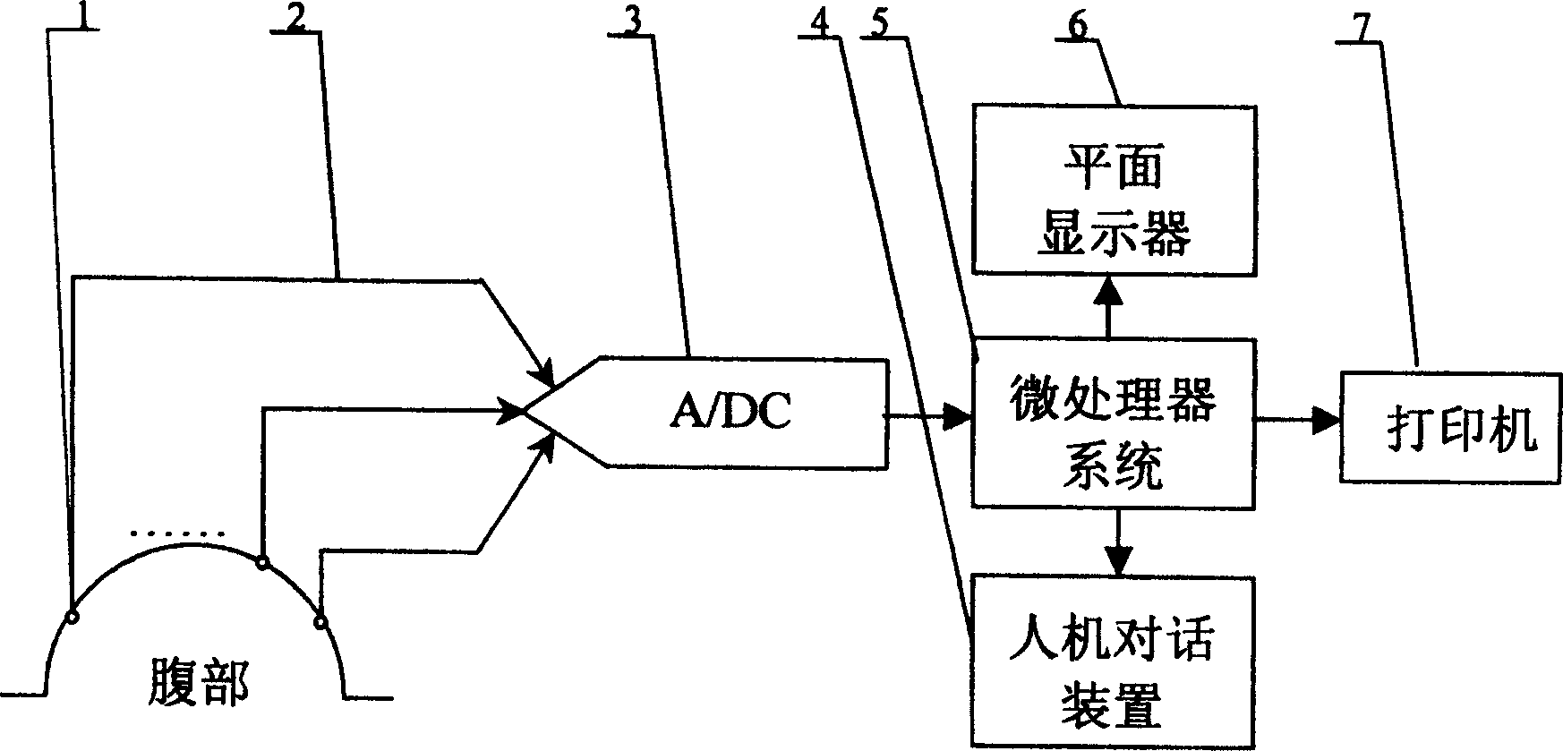
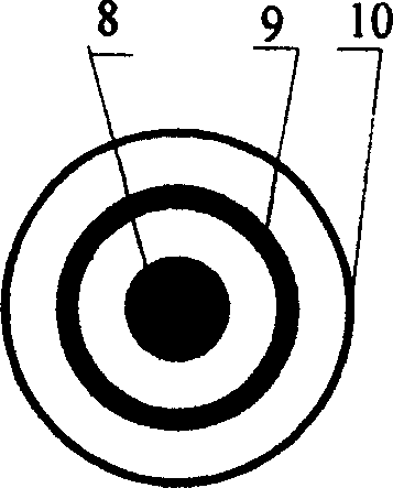
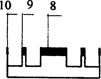
[0022] exist figure 1 Among them, the uterine contraction Laplace topography instrument of the present invention, it is composed of a plurality of Laplace electrodes 1, lead wires 2, analog / digital converter 3, microprocessor system integrating bioelectrical amplifiers and filter circuits 5 and display 6 are connected in turn to form. The microprocessor system 5 is also connected with a printer 7 and a man-machine dialogue device 4, and the microprocessor system 5 can be a single-chip computer or a single-board computer integrated in a chip, or a system computer. The electrode 1 in this embodiment is a plurality of Laplace electrodes integrating bioelectrical amplifiers and filter circuits, and the display 6 is a liquid crystal display. The electrodes are used to pick up the uterine myoelectric Laplace signal of the parturient from the parturient's abdomen, and output it to the analog / digital converter 3 after being amplified and filtered. The analog / digital converter 3 is u...

Embodiment 2

[0026] The difference from the first embodiment is that the display 6 is a CRT monitor, and the man-machine dialogue device 4 is a keyboard and a mouse.

[0027] The working principle of the present invention is: a plurality of Laplace electrodes 1 integrating bioelectrical amplifiers and filter circuits are placed on the abdomen of the puerpera, and the uterine myoelectric Laplace signal picked up by the Laplace electrode 1 from the abdomen of the puerpera passes through After filtering and amplification processing, it is input to the analog / digital converter 3 through the lead line 2, and the converted digital signal is processed by the microprocessor system 5, and the processed results can be displayed by the flat-panel display 6 and printed by the printer 7 respectively , the man-machine dialogue device 4 is used to operate the instrument and input necessary information. The number of electrodes required is 8 to 32, the bandwidth of the integrated filter on the back of the...


PUM


Abstract

The invention discloses a uterine contraction Laplace topography instrument, which is composed of a plurality of Laplace electrodes integrated with bioelectrical amplifiers and filter circuits, an analog-to-digital converter, a microprocessor system and a display connected in sequence, and the micro The processor system is also connected with a man-machine dialogue device. Multiple electrodes are placed in the abdomen of the parturient, and the uterine myoelectric Laplace signal of the parturient is picked up, amplified and filtered, and then input to the analog / digital converter. The converted digital signal is processed by the microprocessor system and can be processed by the plane Display on monitor and print on printer. The uterine contraction Laplace topography instrument of the present invention can not only detect the pacemaker activity of each part of the puerpera's uterus in real time, but also reflect the contraction sequence, phase and direction of each part of the puerpera's uterus, so as to diagnose whether the labor force is normal or not. It provides a scientific basis and provides a new method for avoiding dystocia, guaranteeing the natural delivery of mothers and the safety of mothers and babies.

Description

technical field [0001] The invention belongs to a medical instrument, in particular to an instrument for detecting and displaying the strength and direction of uterine contraction in a puerpera during childbirth. Background technique [0002] There are three main reasons for maternal dystocia during childbirth: abnormal fetus, abnormal birth canal and abnormal labor force. The first two reasons are easy to detect before delivery. For primiparous women, the third reason can only be diagnosed during labor, but there is no instrument or means to detect it. Doctors only rely on experience to judge, without objective indicators, it is difficult to make an accurate diagnosis, and they cannot effectively guarantee the safety of mothers and babies. [0003] Uterine muscle is a kind of smooth muscle, which is different from skeletal muscle, and the frequency and mode of transmission of the two are very different. Although some people use ordinary electr...

Claims


Application Information

Patent Timeline
Patent Type & Authority Patents(China)
IPC IPC(8): A61B5/0488A61B5/04
Inventor 姜苇李刚林凌
Owner TIANJIN UNIV
Learn More