Method for reinforcing edge of medical picture

An edge enhancement and medical image technology, applied in image enhancement, image data processing, instruments, etc., can solve the problem of different edge enhancement effects of images

Inactive Publication Date: 2003-03-26
WUHAN UNIV
View PDF0 Cites 13 Cited by
  • Summary
  • Abstract
  • Description
  • Claims
  • Application Information

AI Technical Summary

Problems solved by technology

These edge enhancement operators have different effects on image edge enhancement due to their different algorithms.

Method used


Image

  • Method for reinforcing edge of medical picture
  • Method for reinforcing edge of medical picture
  • Method for reinforcing edge of medical picture

Examples


Embodiment 1

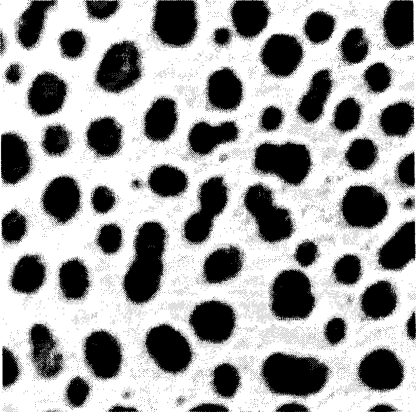
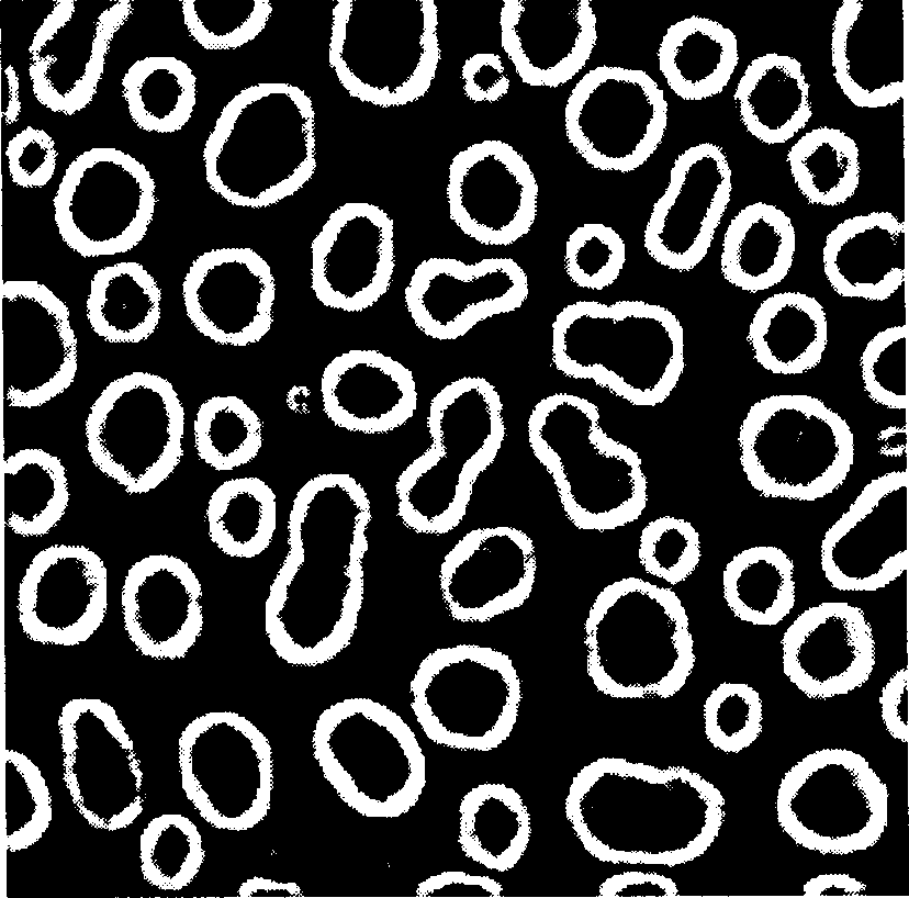
[0041] Embodiment 1: After the original image is input, it is sequentially processed by a rank value filter, a Laplacian filter, an edge detector, a gain unit, and a summation unit to obtain an enhanced medical grayscale image The resulting image of edge features.

Embodiment 2

[0042] Embodiment 2: After the original image is input, it is sequentially processed by a Laplacian filter, an ordinal value filter, an edge detector and a gain unit to obtain an enhanced result image of the edge features of the medical grayscale image.

Embodiment 3

[0043] Embodiment 3: After the original image is input, it is sequentially processed by the first sequence value filter, the second sequence value filter, the Laplacian filter, the edge detector, and the gain unit to obtain an enhanced medical grayscale image The resulting image of edge features. Wherein the sequence value of the first sequence value filter + the sequence value of the second sequence value filter is: N+1 (N is the number of non-zero gray value pixels in the neighborhood).


PUM


Abstract

The edge enhancing operator is obtained by combining the Laplacian filter, the ordered value filter, the edge detector, the gain unit and / or the sum unit. The medical image edge is carried out the digital image processing by using the said operator so as to obtain the clear, continuous, smooth edge features of the medical gray image. With these edge features of the medical image being analyzed, the diagnostic information of the tumor tissue can be picked upand distinguished from the original image captured by various image devices. Also, the tissue structure information, the blood vessel information in the tissue and the boundary information of the gray shade granule related to the relevant area in the image provided can be picked up and distinguished. Thus, the invention provides powerful reference for diagnosing diseases by using the clinical image.

Description

technical field [0001] The invention relates to an edge enhancement method of a medical image. Background technique [0002] With the development of science and technology, new medical imaging equipment representing the latest level of scientific and technological development continues to appear, and the digitization and networking of various medical imaging equipment, resource sharing of medical imaging, and remote consultation are developing rapidly. In modern clinical diagnosis, the diagnosis of almost all kinds of diseases is increasingly relying on various medical imaging equipment and related imaging diagnostic technologies, because various modern and advanced medical imaging equipment can provide doctors with a large amount of rich image information. In modern imaging diagnosis, medical imaging equipment must firstly provide high-definition, high-resolution and high-gray-scale digital images. At the same time, post-processing of digital medical images can display more...

Claims


Application Information

Patent Timeline
IPC IPC(8): G06T5/10
Inventor 李凯扬相艳杨宣东
Owner WUHAN UNIV
Why Eureka
Try Eureka