Registration method of three dimension image

A three-dimensional image and registration technology, which is applied in image analysis, image data processing, and image-to-image conversion, can solve the problems of inability to meet clinical needs and high computational complexity, reduce the possibility of falling into local extremum, and achieve good robustness. Sticky, precisely registered results

Inactive Publication Date: 2007-04-25
SOUTHERN MEDICAL UNIVERSITY
View PDF0 Cites 25 Cited by
  • Summary
  • Abstract
  • Description
  • Claims
  • Application Information

AI Technical Summary

Problems solved by technology

However, the traditional method based on mutual information needs to consider the information of the entire 3D data, and the calculation complexity is large, which cannot meet the clinical needs.

Method used


Image

  • Registration method of three dimension image
  • Registration method of three dimension image
  • Registration method of three dimension image

Examples


Embodiment Construction

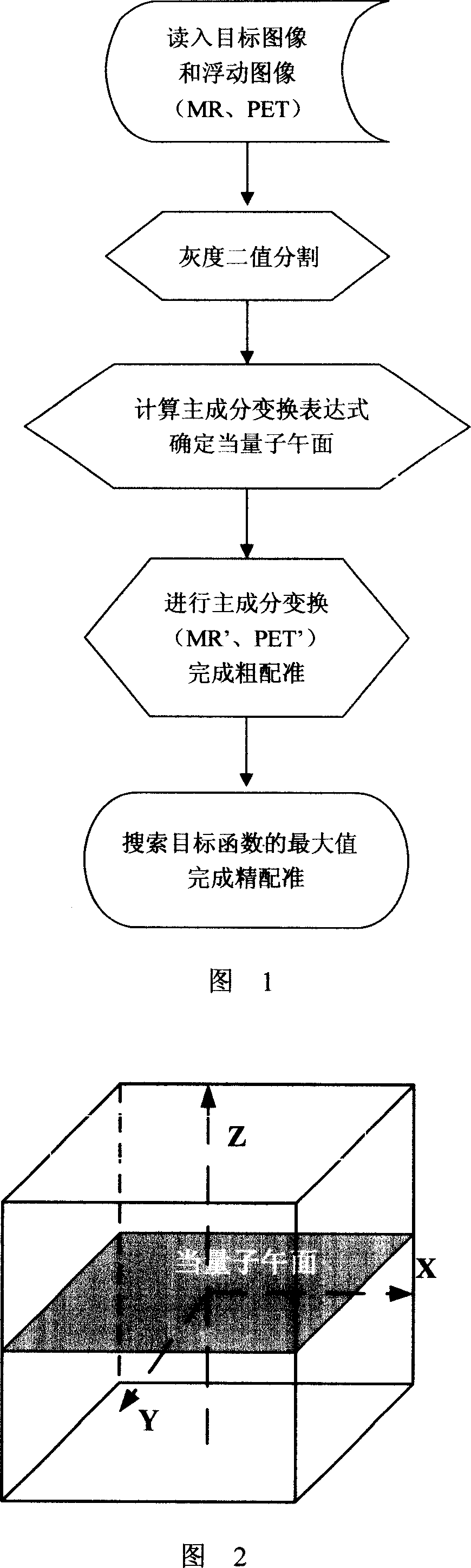
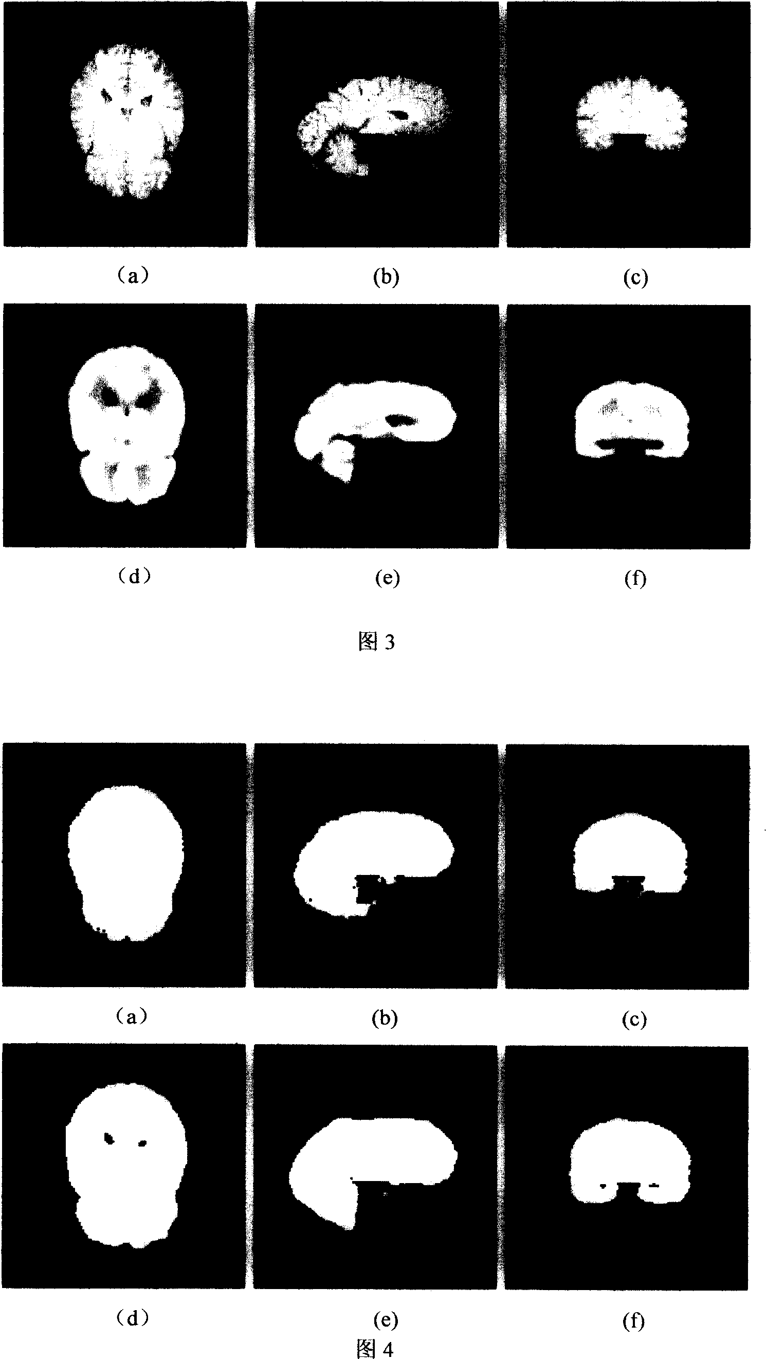
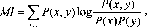
[0020] The working steps of the present invention will be described in detail below in combination with a set of head MR data and PET data (see FIG. 3 ).

[0021] Step 1: Read in MR images and PET images, and use median filtering to preprocess the input images to reduce the impact of noise on registration. By linear transformation (I-I min )×255 / (I max -I min ) Transform the gray value of all pixels into the range of 0-255, where I is the gray value of the image.

[0022] Step 2, use the FCM algorithm to perform binary rough segmentation on the gray value of the MR image to obtain the threshold T0, first take T∈[T 0 -10,T 0 +10], segment the MR image with different T values, and calculate the mutual information between the segmented MR image and the original MR image, and then use the T value when the mutual information is the largest to perform binary fine segmentation on the MR image. Among them, the calculation formula of mutual information is:

[0023] ...


PUM


Abstract

This invention discloses a rapid align method of three dimensional image, which comprises the following steps: a, separately reading aim image and float image; b, separately cutting the image data for grey two values to get vector expression in object; c, computing object mass center and covariance matrix to get main exchange expression and equivalent weight meridional plane; d, using main change to exchange object into original image middle layer to fulfill rough aligning; e, fixing one image as aim image with other as float image.

Description

technical field [0001] The invention relates to a graphic image conversion in an image plane, in particular to a fast registration method of three-dimensional images, which is applicable to the registration of graphic image slices of medicine, meteorological satellites, and aviation. Background technique [0002] Medical image registration refers to seeking a kind of (or a series of) spatial transformation for a medical image to make it consistent with the corresponding points on another image. This consistency means that the same anatomical point on the human body has the same spatial position in the two images, and the registration result should make all the anatomical points on the two images match. Multimodal medical image registration is a hot issue in the field of medical image processing, which is of great significance for clinical diagnosis and treatment. Medical images can provide detailed information such as the size, shape, and spatial relationship of diseased ti...

Claims


Application Information

Patent Timeline
IPC IPC(8): G06T3/00G06T7/00
Inventor 卢振泰陈武凡
Owner SOUTHERN MEDICAL UNIVERSITY
Why Eureka
Try Eureka