
Method and magnetic resonance apparatus for reconstruction of a three-dimensional image data set from data acquired when a noise object distorted the magnetic field in the apparatus

a three-dimensional image and noise object technology, applied in the field of three-dimensional magnetic resonance image reconstruction, can solve the problems of distorted magnetic field, artifacts in the reconstructed image, and the inability to reconstruct diagnostic-quality magnetic resonance images, so as to achieve the effect of improving image quality

Active Publication Date: 2018-11-13
SIEMENS HEALTHCARE GMBH
View PDF24 Cites 0 Cited by
  • Summary
  • Abstract
  • Description
  • Claims
  • Application Information

AI Technical Summary

Benefits of technology

This approach enhances image quality by reducing artifacts and maintaining high signal-to-noise ratio, avoiding the limitations of low-resolution Fourier transforms and signal overshooting, while minimizing the impact of SEMAC correction on undistorted regions.

Problems solved by technology

Reconstructing diagnostic-quality magnetic resonance images becomes problematic when the magnetic field to which the spins in the target region are subjected is distorted during the acquisition of the underlying raw data.
Noise objects cause local distortion of the constant magnetic field, which means that exhibits a local inhomogeneity that influences the otherwise constant magnetic field both the excitation of the nuclear spins and the acquisition of the magnetic resonance signals.
An influence that is problematic when imaging different target slices occurs during the excitation.
This results in artifacts in the reconstructed image.
A problem in this procedure, however, is that the overall measurement time is increased significantly because of the multiplicity of additional phase-encoding steps required per target slice in order to resolve the respective excitation profile of each target slice.
This results in the further problem that the imaging capability of the Fourier transform into the spatial domain (image space) is extremely poor because of the lower resolution.
The “point spread function” causes the acquired magnetic resonance signals to overshoot into adjacent target slices, which means that the image clarity is impaired, or in extreme cases incorrect image information is delivered.
These more subtle limitations on the image quality are hardly noticeable in the distortion area, where the SEMAC correction is extremely effective, wherein image areas predominate that are sufficiently distant from the disturbances caused by the noise object and are thus not subject to any distortions, but where a reduction of the image quality may nevertheless occur as a result of undesired side-effects of the SEMAC reconstruction.

Method used


Image

  • Method and magnetic resonance apparatus for reconstruction of a three-dimensional image data set from data acquired when a noise object distorted the magnetic field in the apparatus
  • Method and magnetic resonance apparatus for reconstruction of a three-dimensional image data set from data acquired when a noise object distorted the magnetic field in the apparatus
  • Method and magnetic resonance apparatus for reconstruction of a three-dimensional image data set from data acquired when a noise object distorted the magnetic field in the apparatus

Examples


Embodiment Construction

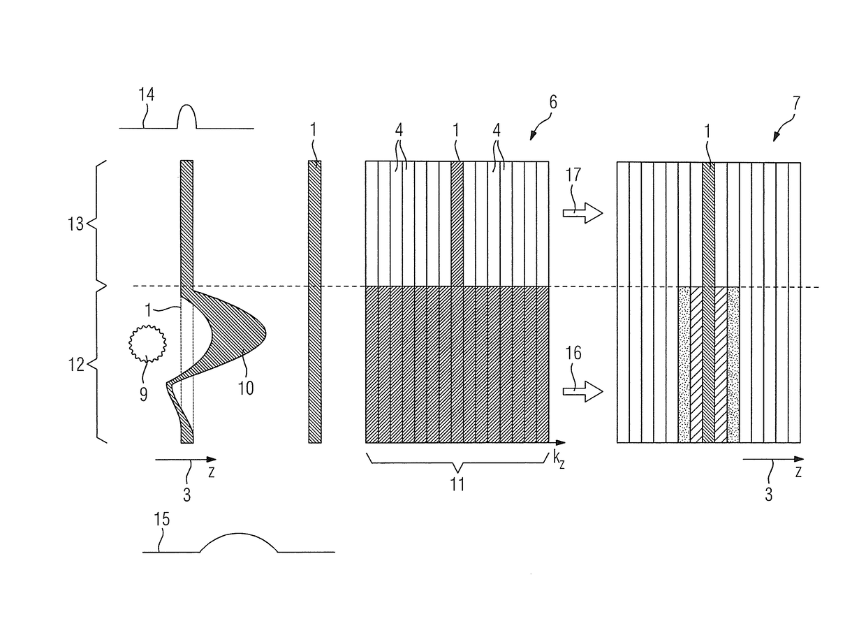
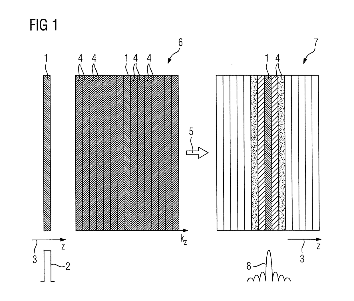
[0032]FIG. 1 shows in detail the problem underlying the present invention with the example of a target slice 1 having no distortions. If one considers the ideal profile 2 of the magnetic resonance signals, in other words slice data, in the slice selection direction 3, which here when using the SEMAC method also corresponds to the supplementary encoding direction, an idealized rectangular function is produced because the entire signal originates from said target slice 1. When using the SEMAC method, however, not only the target slice 1 is scanned as the central partition slice but in further phase-encoding steps along the slice selection direction 3 as a supplementary encoding direction fourteen adjacent partition slices 4 are likewise scanned in the present case, which means that fifteen phase-encoding steps are used in the present case.

[0033]In order to carry out the SEMAC correction, the slice data of the target slice 1 and also of the adjacent partition slices 4 present in the k-...


PUM


Abstract

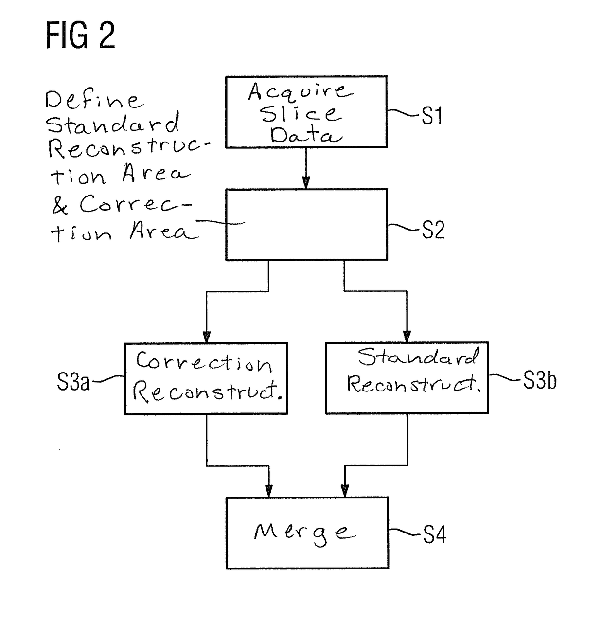
In a method for reconstruction of a three-dimensional image data set from magnetic resonance slice data of a target region acquired in target slices while a noise object distorting the magnetic field is present in the target region, for each target slice to be acquired, in addition to a central partition slice corresponding to the respective target slice, location, multiple partition slices adjacent to the central partition slice are acquired in a supplementary encoding direction perpendicular to the slice plane in multiple phase-encoding steps. A correction area and a standard reconstruction area of the target region are determined on the basis of a distortion criterion, obtained by evaluating the slice data that describes the distortion along the supplementary encoding direction. In the standard reconstruction area, only slice data are used, and in the correction area, slice data of partition slices outside the target slice are assigned to target slices in order to correct the distortion.

Description

BACKGROUND OF THE INVENTION[0001]Field of the Invention[0002]The invention concerns a method and magnetic resonance apparatus for reconstruction of a three-dimensional magnetic resonance image data set from magnetic resonance slice data of a target region acquired in target slices by operation of a magnetic resonance scanner of the apparatus while a noise object distorted the magnetic field in the target region, in particular a metal object.[0003]Description of the Prior Art[0004]Magnetic resonance tomography is an imaging modality in widespread use, with which it is possible to acquire magnetic resonance raw data from a three-dimensional target region inside a patient, by acquiring the data from multiple slices of the three-dimensional target region using 2D imaging for each slice. These different slices of the target region (target slices) normally form a so-called slice package which contains a three-dimensional raw data set, from which a three-dimensional magnetic resonance imag...

Claims


Application Information

Patent Timeline
Patent Type & Authority Patents(United States)
IPC IPC(8): G01R33/565G01R33/48G01R33/56G01R33/54G01R33/483G01R33/561
CPCG01R33/56536G01R33/4828G01R33/5611G01R33/543G01R33/5608G01R33/4835
Inventor NITTKA, MATHIAS
Owner SIEMENS HEALTHCARE GMBH
Learn More