Regularization of images

a technology of regularization and images, applied in image enhancement, tomography, instruments, etc., can solve the problems of limiting the clinical utility of many research tracers, time-consuming, risky for patients, and expensive blood chemistry analysis, etc., and achieve the effect of suppressing ringing artifacts and suppressing artifacts

Active Publication Date: 2019-05-28
THE RES FOUND OF STATE UNIV OF NEW YORK +1
View PDF14 Cites 1 Cited by
  • Summary
  • Abstract
  • Description
  • Claims
  • Application Information

AI Technical Summary

Problems solved by technology

Typically sampling is done via an arterial catheter, a procedure which is costly, risky to patients, time consuming, requires specialist, as well as expensive and error prone blood chemistry analysis.
The requirement of blood sampling limits clinical utility of many research tracers—it is not feasible to conduct tracer blood analysis in a clinic.
Also, many research centers cannot conduct fully quantitative studies using many of the available tracers due to the added expense of blood sampling and analysis.
Iterative reconstruction with point spread function (PSF) modeling improves contrast recovery in positron emission tomography (PET) images, but also introduces ringing artifacts and over enhancement that is contrast and object size dependent.

Method used


Image

  • Regularization of images
  • Regularization of images
  • Regularization of images

Examples


example 1

eighted Total Variation Denoising for Ringing Artifact Suppression in PET Reconstruction Using PSF Modeling

[0147]Existing and commonly used iterative reconstruction techniques in positron emission tomography (PET) provide a flexible framework for modeling the physics and the scanner geometry, yielding greater image contrast, visual quality and noise robustness than analytical reconstruction methods.

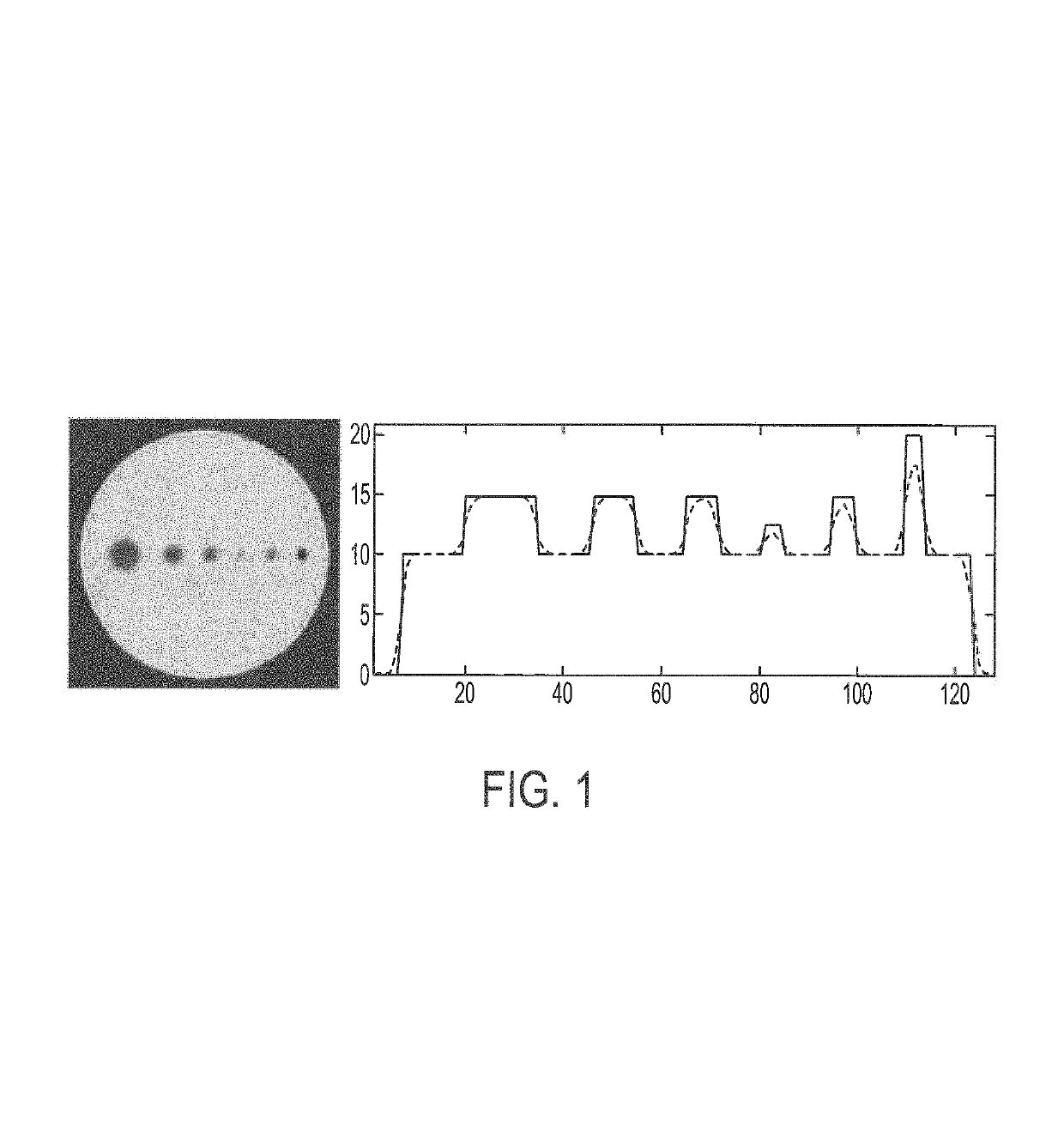
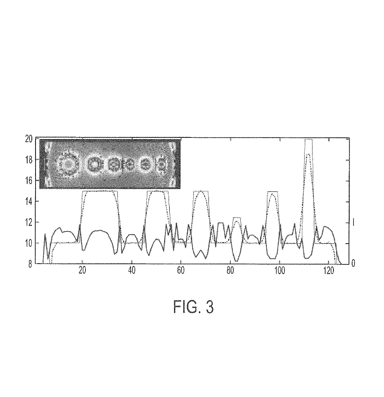
[0148]Modeling and accounting for the physics of the emissions (e.g. positron range) and the detection processes (e.g. crystal scattering or depth of interaction) is possible by measuring and incorporating the detector point spread function (PSF) into the reconstruction process. Such approach has been shown to improve spatial resolution and image contrast (Sureau et al, 2008). Unfortunately, this approach also introduces significant edge artifacts such as over enhancement and Gibbs type of ringing that are both contrast and size dependent (leading to up to 70% overshoot in a cylinder phan...

example 2

eighted Total Variation Denoising for PSF Modeling Artifact Suppression in Pet Reconstruction

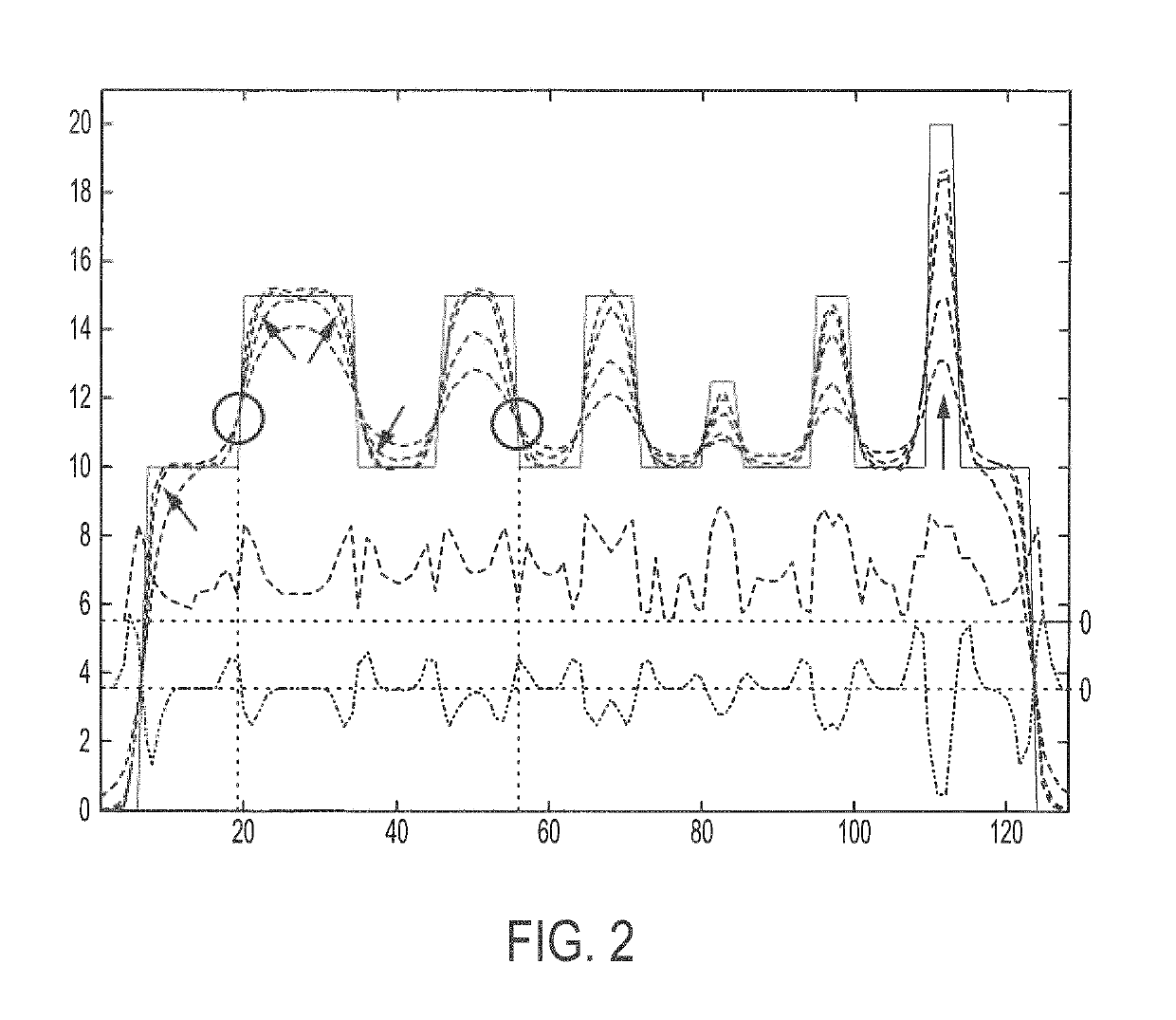
[0185]Incorporating the point spread function (PSF) into the iterative MLEM reconstruction of PET images introduces contrast and size dependent ringing and over enhancement artifacts. As described herein, TV-PSF-MLEM can be used to suppress these artifacts based on the introduction of a locally-weighted total variation regularization within the MLEM reconstruction algorithm. On non-noisy PET measures, the TV spatial weights can be computed based on the point-wise convergence rate of a preliminary MLEM reconstruction, for each voxel. The TV-PSF-MLEM weighting scheme can be extended to noisy measures introducing a noise-independent weighting scheme. As described herein, the performance of TV-PSF-MLEM weighting scheme having a noise-independent weighting scheme can be compared to a state of the art PET denoising method. Results on numerical phantoms show that the TV-PSF-MLEM method offers subst...

example 3

ction Based Resolution Recovery

[0227]A fundamental limitation of PET technology is the image resolution of 8-12 mm, which varies based on scanner and application. In molecular imaging every millimeter matters. Improving the resolution by a single millimeter could open the door to a wide range of applications. More reliable detection of small tumors could translate into earlier diagnosis and better prognosis. The need for quantifying of signal from internal carotid arteries in the brain, that are on the order of 6-8 mm in diameter, has been discussed. Reliable quantitation of carotid signal could facilitate estimation of the arterial input function and lead to less invasive PET quantification. Another application of interest is the quantification of signal from raphe nucleus in the brain, whose five nuclei are spherical structures that are roughly 5-6 mm in diameter. The raphe is a major site of serotonin receptors and regulator of serotonin utilization through the brain. Only a few ...


PUM


Abstract

The present disclosure is directed to iterative regularized reconstruction methods. In certain embodiments, the methods incorporate locally-weighted total variation denoising to suppress artifacts induced by PSF modeling. In certain embodiments, the methods are useful for suppressing ringing artifacts while contrast recovery is maintained. In certain embodiments, the weighting scheme can be extended to noisy measures introducing a noise-independent weighting scheme. The present disclosure is also directed to a method for quantifying radioligand binding in a subject without collecting arterial blood. In certain embodiments, the methods incorporate using imaging data and electronic health records to predict one or more anchors, which are used to generate an aterial input function (AIF) for the radioligand.

Description

[0001]This application is a continuation-in-part of International Application No. PCT / US2015 / 018817 filed Mar. 4, 2015, which claims the benefit of and priority to U.S. provisional patent application Ser. No. 61 / 947,739, filed Mar. 4, 2014, and U.S. provisional patent application Ser. No. 61 / 952,739, filed Mar. 13, 2014, the disclosures of which are hereby incorporated by reference in their entireties for all purposes.[0002]This invention was made with government support under grant MH095338 awarded by the National Institutes of Health. The Government has certain rights in this invention.[0003]This patent disclosure contains material that is subject to copyright protection. The copyright owner has no objection to the facsimile reproduction by anyone of the patent document or the patent disclosure as it appears in the U.S. Patent and Trademark Office patent file or records, but otherwise reserves any and all copyright rights.[0004]All publications, patent applications, patents, and o...

Claims


Application Information

Patent Timeline
Patent Type & Authority Patents(United States)
IPC IPC(8): G06T11/00G06T7/00A61B6/03G06T15/08A61B6/00G16B45/00
CPCG06T7/0012A61B6/037A61B6/481A61B6/501A61B6/5205A61B6/5258G06T11/006G06T15/08G06T11/003G06T2211/424G06T2207/10104G06T2207/10108G06T2207/30016
Inventor MIKHNO, ARTHURANGELINI, ELSA D.LAINE, ANDREW F.OGDEN, TODDPARSEY, RAMINMANN, JOSEPH JOHN
Owner THE RES FOUND OF STATE UNIV OF NEW YORK
Why Eureka
Try Eureka