Haemostatic device

a technology of haemostatic device and sponge, which is applied in the field of haemostatic device, can solve the problems of poor adhesion, poor adhesion of conventional sponge, and inability to conform to the wound in the same manner, so as to increase the surface contact with the wound, facilitate the adhesion, and effectively transmit the effect of the spong

Active Publication Date: 2020-05-19
SELENTUS SCI LTD
View PDF12 Cites 15 Cited by
  • Summary
  • Abstract
  • Description
  • Claims
  • Application Information

AI Technical Summary

Benefits of technology

[0007]Surprisingly, the applicant has found that haemostatic sponges of the invention may significantly improve adhesion to wounds compared to conventional haemostatic sponges. Furthermore, the improved adhesion can be achieved without having to incorporate expensive coagulants, or reactive chemicals. Also, the applicant has found that haemostatic sponges of the invention may adhere readily to partially dry, or lightly bleeding wounds.
[0054]In some embodiments, periodic parallel and straight cuts may be made both along the width of the surface of the sponge and along the length of the surface of the sponge. This may create an array of tissue-contacting elements on the surface of the sponge, each of which has substantially the same cross-sectional shape and cross-sectional area. Varying the cutting positions will allow the number, cross-sectional area and shape of the tissue-contacting elements to be varied.

Problems solved by technology

Conventional foam sponges may not adhere well to tissue because of a poor surface contact.
The porous nature of the material may mean that only a limited amount of the material actually contacts the wound surface, leading to poor adhesion.
Conventional sponges, which have continuous surfaces, may not be able to conform to the wound in the same manner.

Method used


Image

  • Haemostatic device
  • Haemostatic device
  • Haemostatic device

Examples


example 1

on of Surface Modified Gelatin Foam Sponges

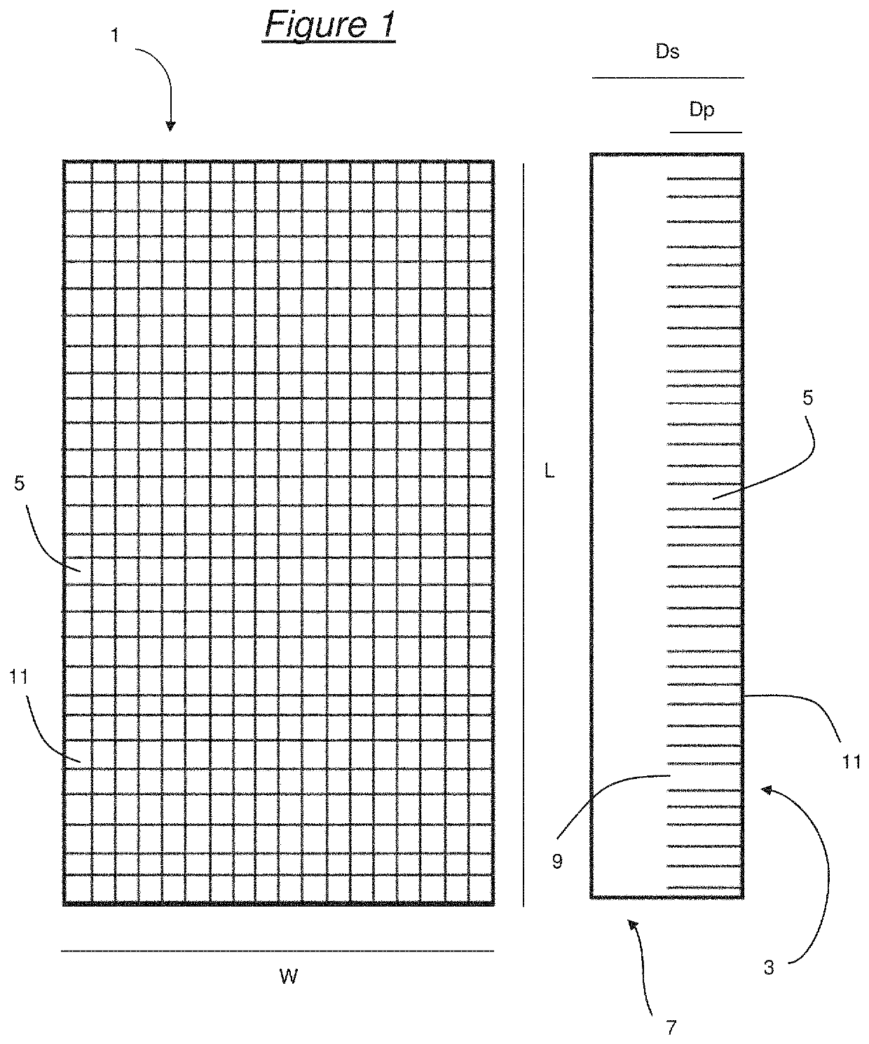
[0116]Sample A: A sponge having the general form shown in FIG. 1 was prepared as follows. A series of incisions were made on a surface of gelatin foam sponges (GelitaSpon by Gelita) using a sharp razor blade (0.1 mm to 0.3 mm width) to create columnar or finger-like tissue-contacting elements on the surface. Incisions were made along the length at 0.5 mm to 2 mm intervals, and to a depth of about 5-8 mm, and a series of widthwise incisions were also made at 0.5 mm to 2 mm intervals at a depth of 5-8 mm. The result was the creation of tissue-contacting elements with substantially rectangular cross-sectional shapes, each having an end face with an area of 0.25 mm2 to 4 mm2. Each sponge had a length of about 30 mm, a width of about 20 mm and a thickness of about 10 mm.

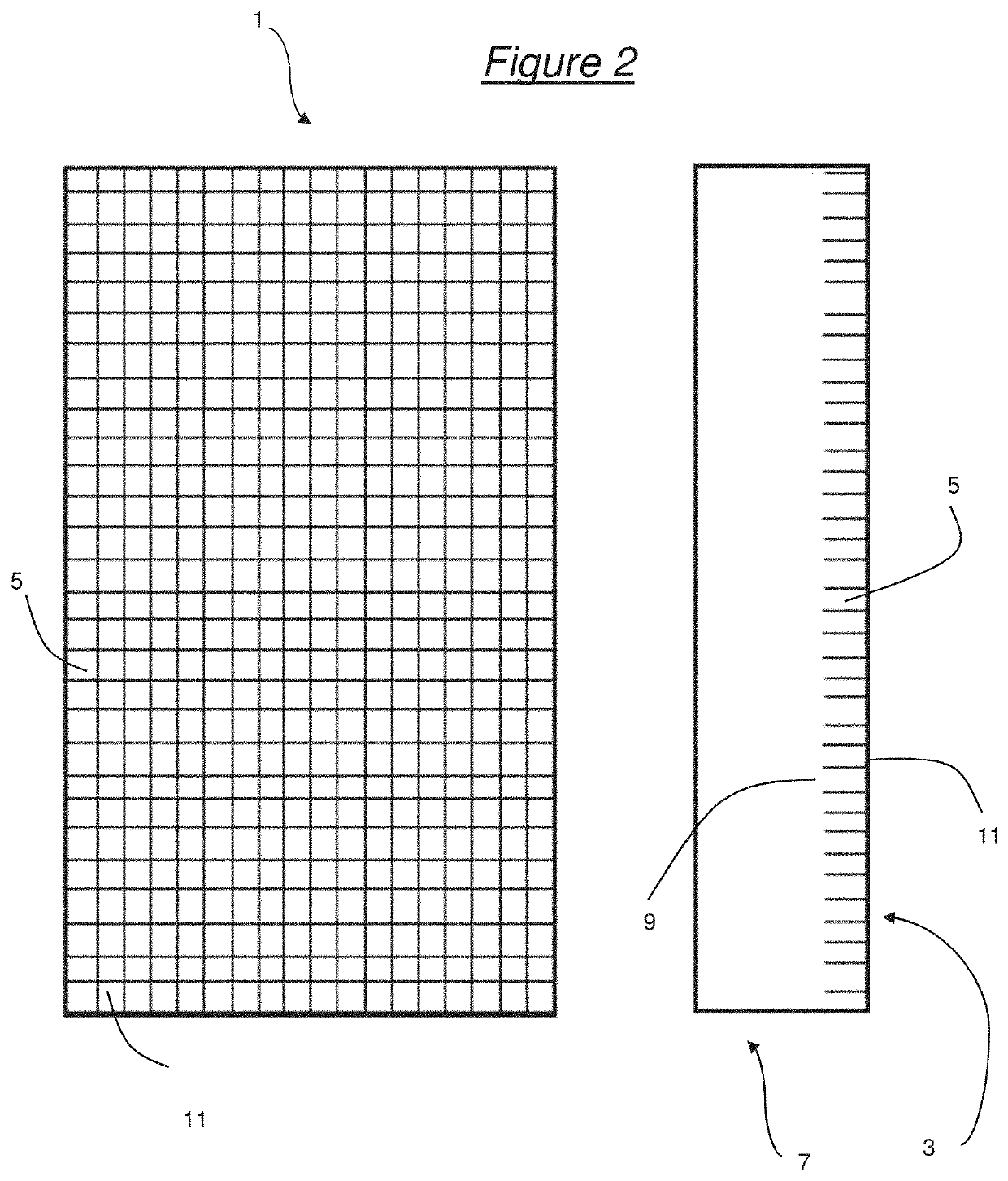
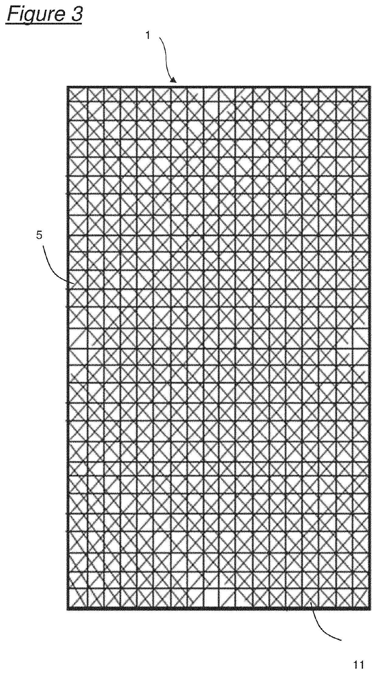
[0117]Sample B: A sponge having the general form shown in FIG. 2 was prepared as follows. A series of incisions were made on a surface of gelatin foam sponges (GelitaSpon by Geli...

example 2

Electron Microscope Images of Surface-Modified Gelatin Foam Sponges

[0122]Scanning Electron Microscope (SEM) images of the surfaces of the some of the samples were taken using a JEOL 6060LV variable pressure scanning electron microscope with a Leica EM SD005 sputter coater.

[0123]FIGS. 5a, 5b and 5c show SEM images of Sample A (plan view) at various magnifications. The face of the tissue-contacting element in FIG. 5b has a surface area of approximately 1.5 mm2.

[0124]FIG. 5d shows a SEM image (×13 magnification) of Sample A (side view) showing tissue-contacting elements with a depth of about 8 mm.

[0125]FIGS. 6a, 6b and 6c show SEM images of Sample B (plan view) at various magnifications.

[0126]FIG. 6d shows an SEM image (×14 magnification) of Sample B (side view) showing tissue-contacting elements with a depth of about 2 mm.

[0127]FIG. 7 shows an SEM image (×13 magnification) of Sample D (plan view).

example 3

dhesion of the Surface-Modified Gelatin Sponges in a Surgical Procedure

[0128]Rabbits (New Zealand White males weighing approximately 3 kg) were prepared and anaesthetised using standard procedures, and the liver uncovered. Biopsy punch holes (5 mm diameter and 4-5 mm deep) were made in the liver lobes, and Samples B and D (control) were placed on the wound and compressed for 1 minute with gauze. Lightly bleeding biopsy wounds were selected such that the sponges would not be fully soaked with blood when placed on the wound. After three minutes the sponge was lifted at one corner with forceps. A further control was also tested: Tachosil (Takeda), a collagen sponge with immobilised thrombin and fibrinogen. Each treatment was replicated three times and the observations were consistent.

Results

[0129]

SpongeImageCommentSample BSee FIGS. 8a and 8bAdhesion is very strong.Lifting the spongecauses the liver to bepulled. The sponge stretchesbut remains attached tothe liver.Sample DSee FIGS. 9a a...


PUM

PropertyMeasurementUnit
depthaaaaaaaaaa
depthaaaaaaaaaa
cross-sectional areaaaaaaaaaaa

Abstract

A bioresorbable haemostatic foam sponge for adhering to a wound. The sponge has a tissue-contacting surface divided into a plurality of closely-spaced tissue contacting elements. Also disclosed are methods for forming the haemostatic sponge and methods of using the sponge.

Description

[0001]This invention relates to haemostatic devices, particularly haemostatic sponges, methods for making such sponges and their uses.BACKGROUND TO THE INVENTION[0002]Foam sponges manufactured from materials such as gelatin or collagen are commonly used to control bleeding which occurs during surgery. These products range in size, typically from 10 mm×10 mm up to 80 mm×120 mm, with a thickness ranging typically from 2 mm to 10 mm. They are made from bioresorbable materials so they can be left in situ in, or on, the body. Commercially available products include Surgifoam (Ethicon), Gelfoam (Pfizer), Ultrafoam (Bard-Davol) and GelitaSpon (Gelita).[0003]Foam sponges control bleeding by providing a porous matrix that adsorbs blood, and provides a surface that promotes clotting. Clotting factors may also be concentrated as the matrix of the sponge absorbs fluid. These products are reasonably effective and widely used in surgery. They may be used dry, or wetted with saline, or to increase...

Claims


Application Information

Patent Timeline
Patent Type & Authority Patents(United States)
IPC IPC(8): A61L31/16A61L15/42A61L15/64A61F13/00A61L31/14A61L15/32
CPCA61L31/148A61L31/16A61F13/00021A61L15/64A61L31/146A61L15/425A61L15/32A61F2013/00463A61L2400/04A61L2300/418A61F13/15A61L15/42A61F13/45A61F13/00063A61F13/36A61L15/44A61L24/0015A61L24/0036A61L24/106
Inventor NICHOLS, JOHN BENJAMIN
Owner SELENTUS SCI LTD
Why Eureka
Try Eureka