
Systems and methods for automated analysis of heterotopic ossification in 3D images

a technology of heterotopic ossification and 3d image, applied in the field of image processing and analysis, can solve the problems of complex image analysis of such diseases, difficult and robust image segmentation, and difficult segmentation, and achieve the effect of facilitating streamlined quantitative analysis of ho formation

Active Publication Date: 2020-10-27
PERKINELMER HEALTH SCIENCES INC
View PDF19 Cites 0 Cited by
  • Summary
  • Abstract
  • Description
  • Claims
  • Application Information

AI Technical Summary

Benefits of technology

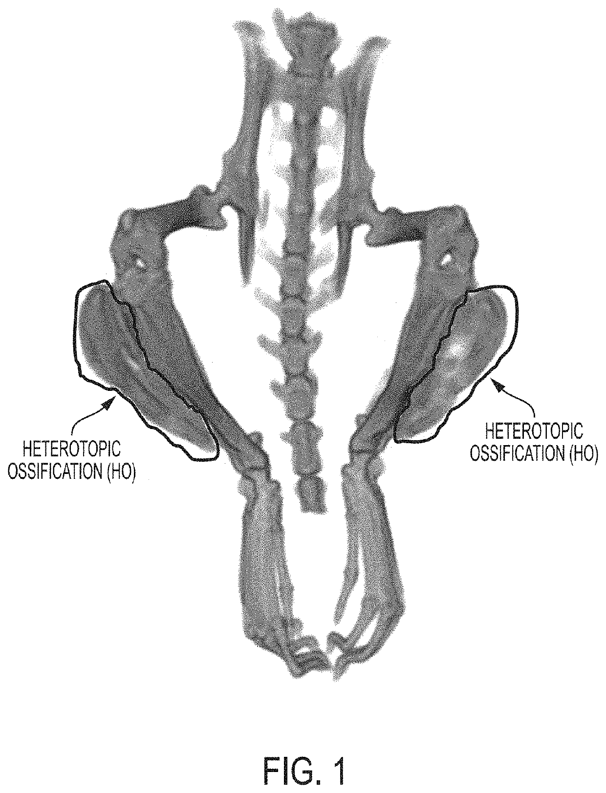
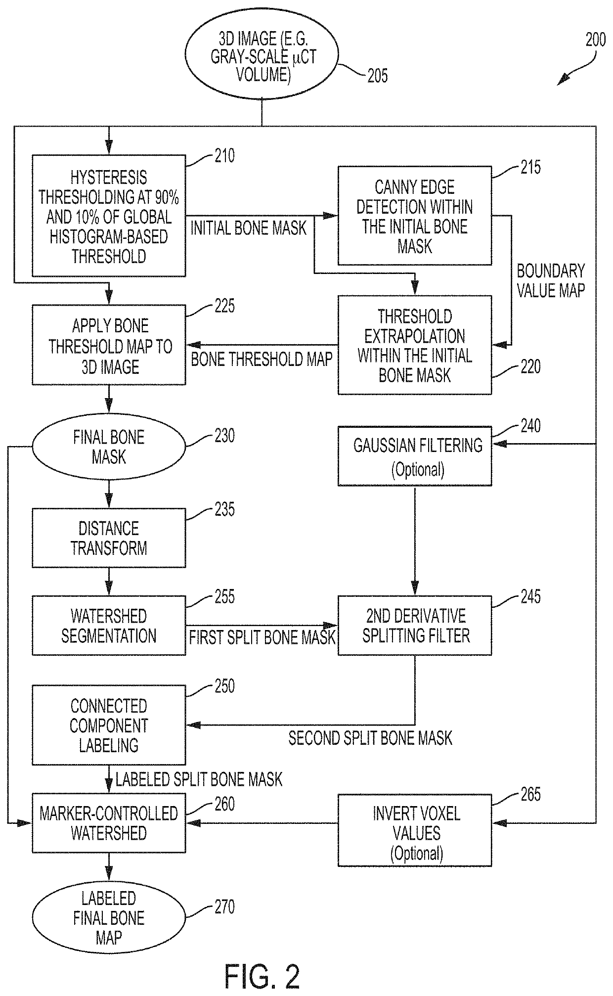
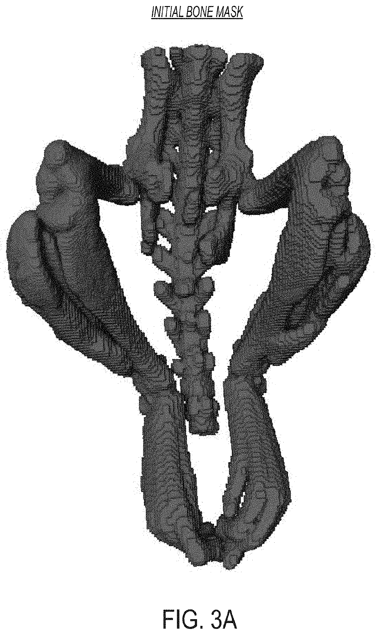
[0010]The approaches described herein utilize a hybrid thresholding approach as well as multiple splitting operations. In certain embodiments, the hybrid thresholding approach and multiple splitting operations account for unique image processing challenges posed by the physical characteristics of HO and resulting characteristics of image regions representing HO. The approaches described herein thereby provide for detection and segmentation of regions in an image corresponding to normal skeleton and HO in a robust and computationally efficient fashion. The detection and segmentation approaches described herein thereby facilitate streamlined quantitative analysis of HO formation, including analysis of morphometric attributes, density, and structural parameters. For example, automated quantification of HO volume in longitudinal studies provides insight into efficacy and kinetics of different treatments. Accordingly, by allowing improved quantitative analysis of HO in this manner, the systems and methods described herein provide a valuable tool for assessing disease state and / or progression in a subject and for assessing treatment efficacy.

Problems solved by technology

Where different structures of interest are similar in nature and / or found in close proximity to each other, accurate and robust image segmentation can be challenging.
These segmentation challenges are also encountered in the analysis of images of certain diseases.
This makes image analysis of such diseases more complicated.

Method used


Image

  • Systems and methods for automated analysis of heterotopic ossification in 3D images
  • Systems and methods for automated analysis of heterotopic ossification in 3D images
  • Systems and methods for automated analysis of heterotopic ossification in 3D images

Examples


Embodiment Construction

[0068]It is contemplated that systems, architectures, devices, methods, and processes of the claimed invention encompass variations and adaptations developed using information from the embodiments described herein. Adaptation and / or modification of the systems, architectures, devices, methods, and processes described herein may be performed, as contemplated by this description.

[0069]Throughout the description, where articles, devices, systems, and architectures are described as having, including, or comprising specific components, or where processes and methods are described as having, including, or comprising specific steps, it is contemplated that, additionally, there are articles, devices, systems, and architectures of the present invention that consist essentially of, or consist of, the recited components, and that there are processes and methods according to the present invention that consist essentially of, or consist of, the recited processing steps.

[0070]It should be underst...


PUM


Abstract

Presented herein are systems and methods that facilitate automated segmentation of 3D images of subjects to distinguish between regions of heterotopic ossification (HO) normal skeleton, and soft tissue. In certain embodiments, the methods identify discrete, differentiable regions of a 3D image of subject (e.g., a CT or microCT image) that may then be either manually or automatically classified as either HO or normal skeleton.

Description

FIELD OF THE INVENTION[0001]This invention relates generally to methods and systems of image processing and analysis. More particularly, in certain embodiments, the invention relates to automatic splitting and segmentation of heterotopic ossification from an anatomical image of a small subject (e.g., small animal, small mammal), e.g., captured with a computed tomography (CT) scanner.BACKGROUND OF THE INVENTION[0002]There is a wide array of technologies directed to in vivo imaging of mammals—for example, bioluminescence, fluorescence, tomography, and multimodal imaging technologies. In vivo imaging of small mammals is performed by a large community of investigators in various fields, e.g., oncology, infectious disease, and drug discovery.[0003]In vivo micro computed tomography (hereafter, “microCT”) imaging, is an x-ray-based technology that can image tissues, organs, and non-organic structures with high resolution, although higher-throughput imaging may make beneficial use of lower ...

Claims


Application Information

Patent Timeline
Patent Type & Authority Patents(United States)
IPC IPC(8): G06T7/187G06F3/0484G06K9/62G06T5/20G06T7/00A61B6/00G06T7/136G06T7/11G06T7/13A61B6/03
CPCA61B6/505G06T5/20G06K9/6202A61B6/5217G06T7/11A61B6/032G06T7/136G06T7/187G06T7/13G06F3/04842G06T7/0012G06T2207/10028G06T2207/30008G06T2207/20012G06T2207/20152G06T2207/10081G06V10/751
Inventor BEHROOZ, ALI
Owner PERKINELMER HEALTH SCIENCES INC
Learn More