
Exosomes sourced from granulocytic myeloid-derived suppressor cells and application thereof

a technology of exosomes and myeloids, applied in the field of cell biology, molecular biology and clinical application, can solve the problems of high yield and short whole process time, and achieve the effects of convenient large-scale extraction, short time-consuming, and easy preservation

Active Publication Date: 2021-10-26
JIANGSU UNIV
View PDF8 Cites 0 Cited by
  • Summary
  • Abstract
  • Description
  • Claims
  • Application Information

AI Technical Summary

Benefits of technology

[0009]G-MDSC exo of the present invention are membranous vesicles composed of derived cell membrane lipid components, which has protection effects on their carried bioactive ingredients. G-MDSC exo can play immunosuppressive roles similar to G-MDSCs, and the exosomes have many advantages that G-MDSCs do not have, they are composite informosomes of subcellular structure for intercellular information transmission.
[0021]It is a further advantage that G-MDSC exo inhibits CD4+ T cell proliferation and promotes Treg expansion, it provides a new concept for the treatment of autoimmune diseases.

Problems solved by technology

The whole process is short time-consuming, high yield.

Method used


Image

  • Exosomes sourced from granulocytic myeloid-derived suppressor cells and application thereof
  • Exosomes sourced from granulocytic myeloid-derived suppressor cells and application thereof
  • Exosomes sourced from granulocytic myeloid-derived suppressor cells and application thereof

Examples


example 1

orting and the Preparation of Culture Supernatant

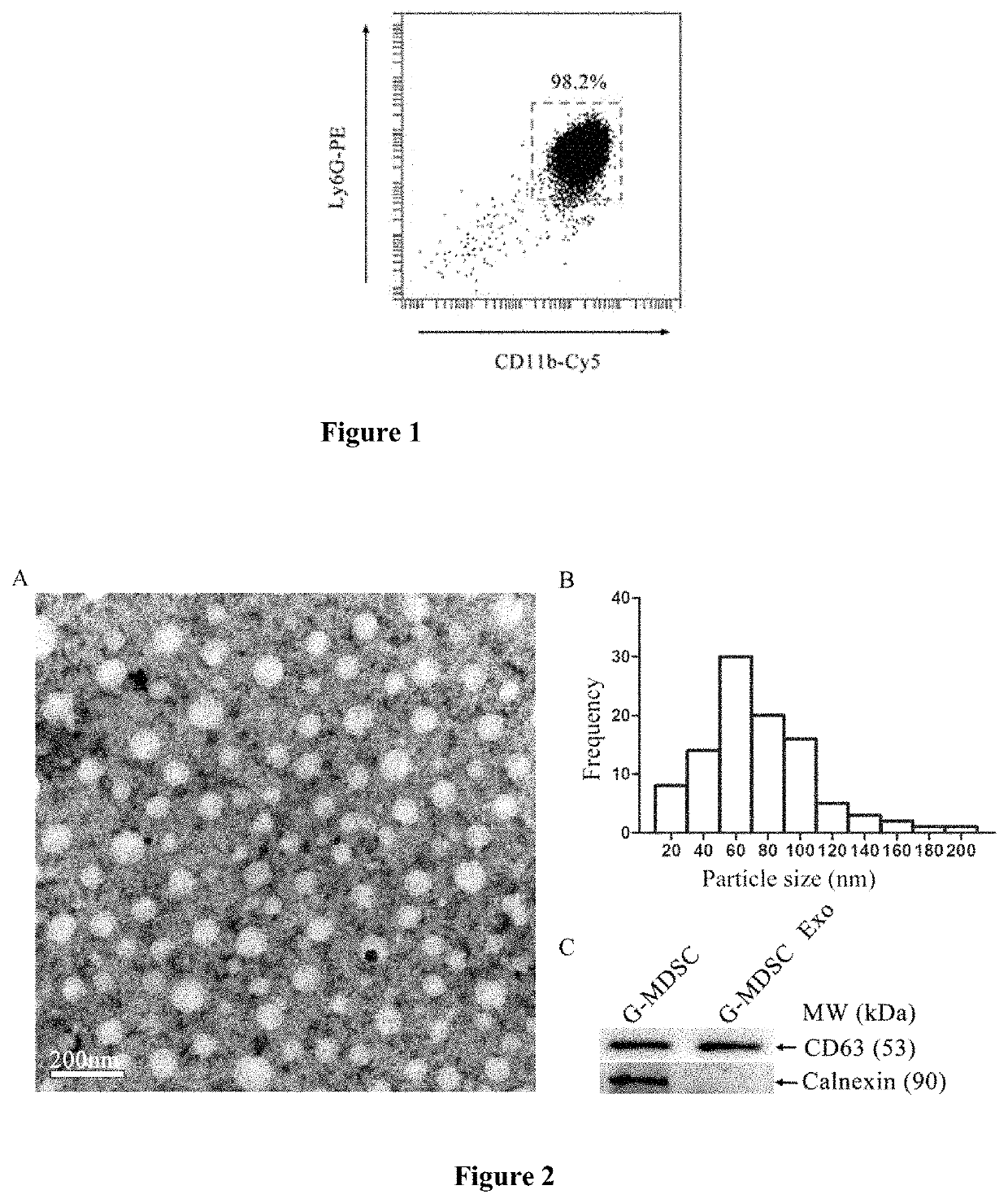
[0030](1) The model of tumor-bearing mouse was established with the Lewis lung adenocarcinoma cell line (LLC): Lewis lung adenocarcinoma cells were cultivated in an incubator at 37° C. and 5% CO2 in the medium (DMED with pH 7.2 and 10% fetal bovine serum). When the cell density is about 85% of the petri dish bottom area, cells are digested with 0.25% trypsin. Male 6-8 w C57BL / 6 mouse were subcutaneously injected at the right side of the abdomen in a dose of 3.0×10⁶ cells per mouse in logarithmic growth phase. The growth of tumors was observed after tumor planting.

[0031](2) Establishment of CIA model: An equal volume of bovine collagen type II (C II) and complete Freund's adjuvant are mixed at a ratio of 1:1 and grind until the mixture is completely emulsified, The degree is that dropping the emulsion into water and it is not loose (operation in ice-bath). Emulsified C II (0.1 mL / mouse) was injected intradermally in the base of the tai...

example 2

on of G-MDSC Exo and Detecting of Protein Concentration

[0036](1) The harvested G-MDSCs supernatant was centrifuged at 4° C., 1000 g for 30 min, the supernatant was collected and centrifuged at 4° C., 10000 g for 30 min. The supernatant was transferred to an ultrafiltration centrifugal tube with MWCO 100 kDa and was centrifuged at 1500 g for 30 min, and the concentrated liquid in the tube was collected.

[0037](2) G-MDSC exo was extracted by ExoQuick-TC™ Exosome Kit purchased from SBI as follows: The concentrated liquid collected in step (1) was mixed with ExoQuick-TC™ Exosome reagent (v / v=5:1), the mixture was vibrated and followed with a standing at 4° C. for more than 12 h and centrifuged at 4° C., 1000 g for 30 min, the precipitate was G-MDSC exo. G-MDSC exo was dissolved in PBS, dispensed to EP tube, and stored at −80° C. for subsequent testing.

[0038](3) Determine the protein concentration of G-MDSC exo by using the BCA Protein Assay Kit: The G-MDSC exo suspension was mixed with t...

example 3

ation of G-MDSC Exo

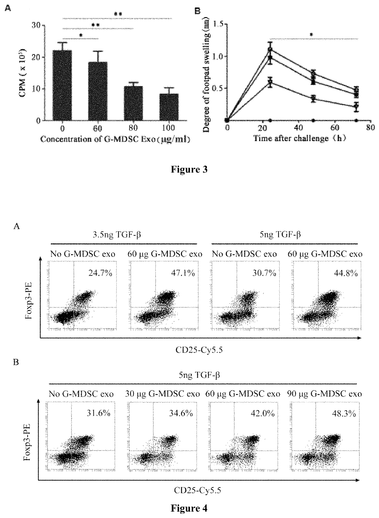
[0039](1) Observing the morphology of G-MDSC exo through transmission electron microscopic: 20 μL of G-MDSC exo suspension were dropped on a 3 mm diameter of sample loading copper mesh, and rest for 2 minutes at room temperature; using filter paper to sip up the liquid gently, and drop 2% of phosphotungstic acid at pH 6.8 on the copper mesh and negatively staining for 1 min, using filter paper sip up the dye liquid and dried under incandescent light bulb. G-MDSC exo were observed as circular or elliptic micro-capsule structure having envelope by transmission electron microscopy, and the intracavity has low electron density components with particle size of 30-150 nm. The result is showed in FIGS. 2A and 2B. FIG. 2A shows the morphology of G-MDSC exo, the G-MDSC exo were observed as circular or elliptic micro-capsule structure having complete envelope by transmission electron microscopy, and the intracavity has low electron density components. FIG. 2B shows the freq...


PUM

PropertyMeasurementUnit
diameteraaaaaaaaaa
diametersaaaaaaaaaa
pHaaaaaaaaaa

Abstract

Provided are exosomes sourced from a granulocyte myeloid-derived suppressor cell and an application thereof. The exosomes are named as G-MDSC exo. Also provided is a use of the exosomes in preparing a drug used for suppressing autoimmune diseases. The G-MDSC exo can effectively suppress proliferation of CD4+T cells in vitro, promote induced proliferation of T regulatory (Treg) cells, alleviate foot swelling of model mice having delayed-type hypersensitivity, and suppress attacks of inflammatory bowel disease (IBD) and collagen-induced arthritis (CIA) of the mice.

Description

FIELD OF INVENTION[0001]This invention relates to granulocytic-like myeloid-derived suppressor cells derived exosomes (G-MDSC exo) and application thereof, which belongs to the fields of cell biology, molecular biology and clinical application. Specifically, the present invention relates to the preparation of G-MDSC exo from spleens of tumor-bearing mouse, the research of biological function and its application in treatment of murine autoimmune diseases.BACKGROUND OF THE INVENTION[0002]Myeloid-derived suppressor cells (MDSCs) are myeloid-derived heterogeneous cells that proliferate greatly under pathological cases of tumor, inflammation and pathogen infection (Gabrilovich D I, Nagaraj S. Myeloid-derived suppressor cells as regulators of the immune system. Nat Rev Immunol. 2009; 9(3):162-74.). MDSCs of mouse are Gr-1 (consisting of Ly6G and Ly6C markers) and CD11b double-positive cells. Two subtypes of MDSCs can be divided into by cell morphology and the expression level of Ly6G and ...

Claims


Application Information

Patent Timeline
Patent Type & Authority Patents(United States)
IPC IPC(8): A61K35/15C12N5/09C12N5/0783
CPCA61K35/15C12N5/0637C12N5/0693A61P19/02A61P29/00A61P37/02A61P37/08
Inventor WANG, SHENGJUNWANG, YUNGANGTIAN, JIERUI, KEMA, JIEMA, BINTANG, XINYIXU, HUAXI
Owner JIANGSU UNIV
Learn More