X-ray apparatus having a composite field of view

a composite field and x-ray technology, applied in the field of x-ray apparatuses with composite field of view, can solve the problems of increasing the dose absorbed by the patient, inefficiency in the radiological department,

Active Publication Date: 2022-07-05
KONINKLJIJKE PHILIPS NV
View PDF6 Cites 0 Cited by
  • Summary
  • Abstract
  • Description
  • Claims
  • Application Information

AI Technical Summary

Benefits of technology

[0006]Accordingly, an apparatus may be provided capable of controlling an X-ray imager which identifies an acquisition deficiency in a first X-ray image. Settings are generated which can be used to adjust the field of view of the X-ray imager, providing a further X-ray image which completes the imaging of the region of interest. The updated field of view may, for example, be smaller in area than the initial field of view. In this case, a patient would be subjected, upon the retake of the X-ray image, to a lower dose of X-ray radiation, compared to the first X-ray image exposure. Such an apparatus enables a reduction in the total X-ray dose delivered to a patient.
[0016]Accordingly, the updated field of view state provides an updated field of view which is as small as possible, because it does not overlap with the field of view of the first X-ray image data. Thus, exposure of the patient to unnecessary X-ray radiation may be further reduced.
[0024]Accordingly, initial or updated fields of view may be defined using the position of a patient in the field of view as captured by, for example, a camera. This enables the initial field of view to be set accurately, as well as the updated field of view, further reducing unnecessary X-ray exposure of the patient.
[0026]Accordingly, the apparatus can be configured to wait for an operator command before activating an X-ray source which performs the second exposure of the patient, thus enhancing the safety of the apparatus.
[0051]Thus, it is a basic idea of the present specification to provide an automated method for robustly checking an initial X-ray image for problems with the field of view, automatically resetting the field of view to enable a second acquisition of anatomical regions not exposed within the initial exposure, and thereby to complete the full field of view using an image combination method. In this way, erroneous initial exposures may be utilized to complete a second exposure which has a smaller field of view, enabling a reduction in radiation exposure of a patient, compared to the radiation exposure that would be needed to undertake two complete images.

Problems solved by technology

Such occurrences are undesirable, leading to inefficiency in radiological departments, and leading to an increase in the X-ray dose absorbed by a patient.

Method used


Image

  • X-ray apparatus having a composite field of view
  • X-ray apparatus having a composite field of view
  • X-ray apparatus having a composite field of view

Examples


Embodiment Construction

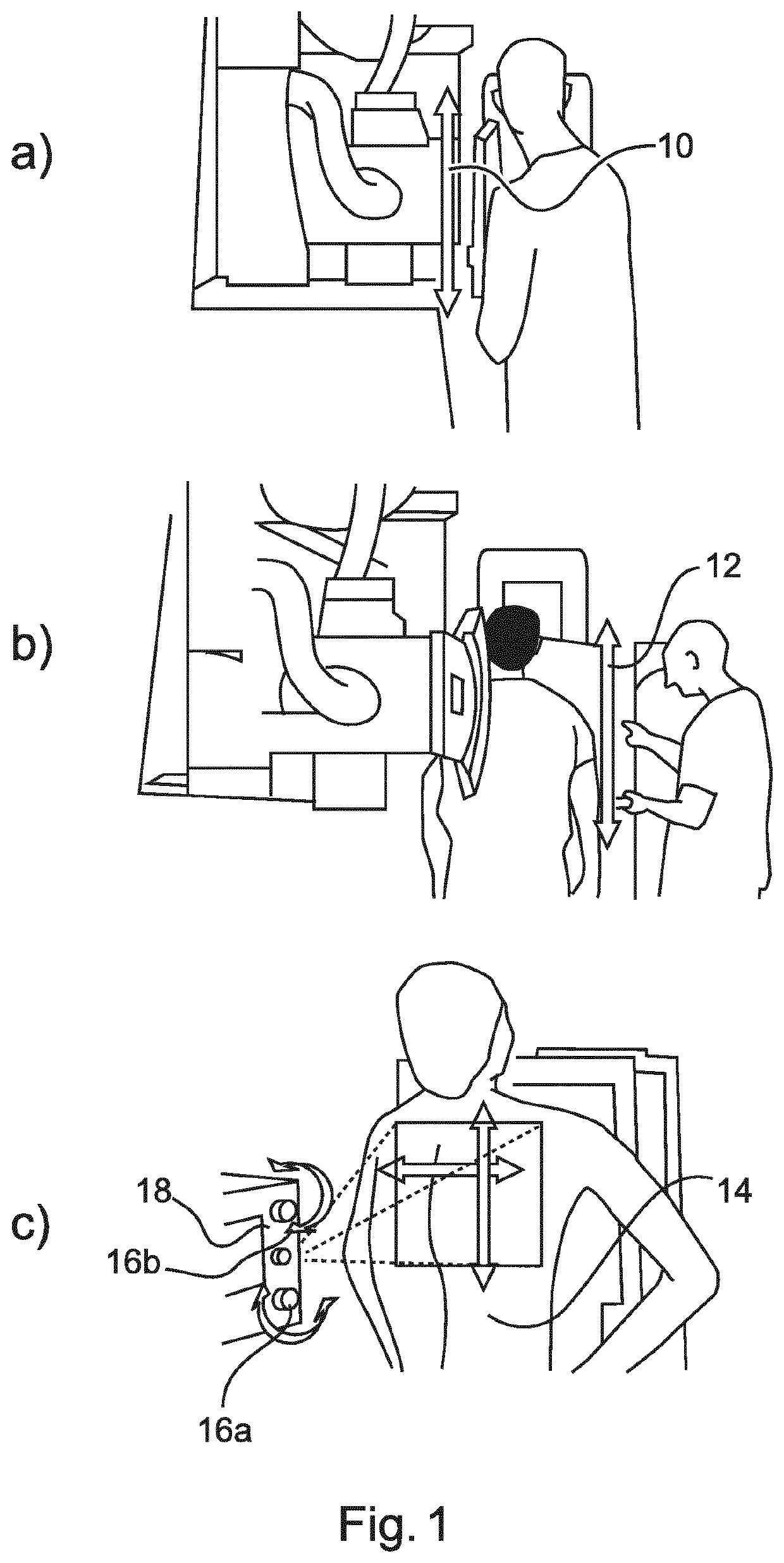
[0063]Chest radiography is the most commonly performed clinical imaging examination, and plays an important role in detecting and diagnosing many diseases of the thoracic anatomy. The image quality is dependent on a wide range of specific individual factors such as the inclusion of the appropriate anatomy within the field of view, the contrast of structures of interest with respect to the background signal, as well as several aspects of the positioning of the patient's thorax with respect to the X-ray acquisition equipment.

[0064]The task of setting the field of view (FOV) for an exposure is conventionally performed by a radiographer. The patient is initially positioned in a region of interest in front of an X-ray detector. Then, a visible light, shining from within the tube-head of the X-ray equipment, and matching to the field of the X-ray radiation pattern, is used to establish the field of view on a patient's body. The height of the tube-head may firstly be varied, and then the h...


PUM


Abstract

Medical radiography requires specialist control of radiography equipment to achieve good imaging results. Typical errors that can occur consist of an inappropriate field of view being accidentally applied. This results in a “cropping effect” in which portions of the region of interest of a patient which would be of clinical use are omitted from the image. Conventionally, the only solution is to re-take the entire image with a more appropriate (and inevitably larger) field of view selected. This is undesirable, because it might require recall of the patient from another location, and the patient will be subject to two exposures, thus undesirably increasing their X-ray dosage. The present application proposes to use an anatomical atlas to analyse an X-ray image output from an initial exposure, in particular to assess whether significant anatomical elements are missing from the image. If elements are missing, the field of view of the X-ray imager is recalibrated, using collimation or pan / tilt adjustments, for example. A subsequent X-ray image is obtained, and combined with the initial image, to provide an output image. Because only a small area of the region of interest may subsequently need to be exposed, a smaller additional dose results.

Description

FIELD OF THE INVENTION[0001]This invention relates generally to an apparatus for obtaining an X-ray image having a composite field of view. Also discussed is an X-ray imaging system, a method for obtaining an X-ray image having a composite field of view, a computer program element, and a computer-readable medium.BACKGROUND OF THE INVENTION[0002]The configuration of the field of view (FOV) for radiographic exposures such as lung radiographs, is a non-trivial task requiring the attention of a trained radiographer to operate an X-ray imager. A field of view which is set erroneously before an exposure means that a subsequent full X-ray image must be taken, because important anatomical detail could be missing from the first image. A patient might even need to be recalled for a second visit. Such occurrences are undesirable, leading to inefficiency in radiological departments, and leading to an increase in the X-ray dose absorbed by a patient. US 2015 / 0228071 A1 discusses an apparatus and...

Claims


Application Information

Patent Timeline
Patent Type & Authority Patents(United States)
IPC IPC(8): A61B6/00G06T7/00
CPCA61B6/5241G06T7/0014G06T2207/10116G06T2207/20221G06T2207/30061G06T2207/30168A61B6/06A61B6/50A61B6/545
Inventor YOUNG, STEWARTBYSTROV, DANIELVON BERG, JENS
Owner KONINKLJIJKE PHILIPS NV
Why Eureka
Try Eureka