Segmentation of the cardiac region in CT images

a segmentation and cardiac region technology, applied in image analysis, image enhancement, instruments, etc., can solve problems such as vessel constrictions

Active Publication Date: 2022-07-12
BAYER AG
View PDF6 Cites 0 Cited by
  • Summary
  • Abstract
  • Description
  • Claims
  • Application Information

AI Technical Summary

Benefits of technology

[0037]According to some embodiments, a contrast agent was intravenously administered beforehand to the person, also referred to as patient in this description, in order to enhance the contrast between, firstly, the blood vessels, through which contrast agent flows, and the ventricles in the cardiac region and, secondly, the bones and the remaining tissue.
[0079]What is meant by “automated” is that no human intervention at all is required. Thus, according to some embodiments of the disclosure, a computer program is installed on a computer system which has access to computed tomography images of the cardiac region of patients and which segments the images in an automated manner without a person having to input parameters or to monitor the process.

Problems solved by technology

These clog and constrict the vessels.

Method used


Image

  • Segmentation of the cardiac region in CT images

Examples


Embodiment Construction

[0084]The disclosure is described below with reference to figures, without wishing to restrict the invention to the features or combinations of features that are shown in the figures, where:

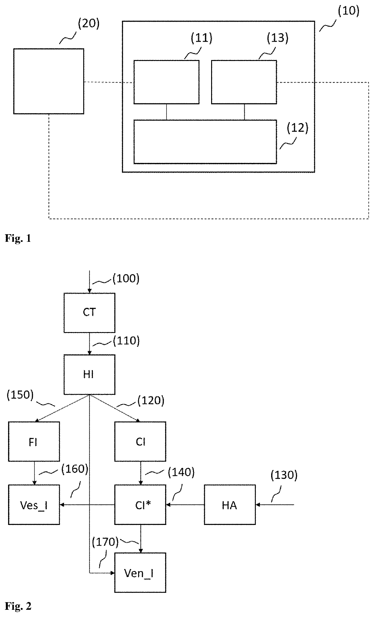
[0085]FIG. 1 shows an exemplary schematic of a computer system according to some embodiments. The computer system (10) comprises a receiving unit (11), a control and calculation unit (12) and an output unit (13).

[0086]Via the receiving unit (11), the computer system (10) receives CT images of the cardiac region of patients. In this description, the term “receive” is also intended to encompass the term “retrieve or read”. According to some embodiments, the CT images can, for example, be read from a data storage medium (20), which can be a component of the computer system according to the disclosure or can be connected thereto via a network (depicted by the dashed lines).

[0087]From the receiving unit (11), the CT images are forwarded to the control and calculation unit (12), which undertakes the au...


PUM


Abstract

The present disclosure pertains to the analysis of the cardiac region in CT images. Provided herein are a method, a computer system and a computer program product for the segmentation of the cardiac region in CT images.

Description

CROSS REFERENCE TO RELATED APPLICATIONS[0001]This application claims priority and benefit to European Application No.: 19204270.3, filed Oct. 21, 2019.FIELD OF THE DISCLOSURE[0002]The present disclosure deals with the analysis of the cardiac region in CT images. Subjects of the disclosure are a method, a computer system and a computer program product for the segmentation of the cardiac region in CT images.BACKGROUND OF THE DISCLOSURE[0003]Computed tomography (CT) is an X-ray method which depicts the human body in cross-sectional images (sectional imaging method). Compared to a conventional X-ray image, on which usually only coarse structures and bones are identifiable, CT images also capture in detail soft tissues with small differences in contrast. An X-ray tube generates a so-called X-ray fan beam, which penetrates the body and is attenuated to varying degrees within the body owing to the various structures, such as organs and bones. The receiving detectors opposite the X-ray emit...

Claims


Application Information

Patent Timeline
Patent Type & Authority Patents(United States)
IPC IPC(8): G06K9/00G06T7/11G06V10/40G06V10/46
CPCG06T7/11G06V10/40G06T2207/10081G06T2207/30048G06T2207/30101G06V10/467G06T7/136G06T7/155G06T7/187G06T2207/20128
Inventor FOIS, FRANCOLIAO, WEIRECHSTEINER, DANIEL
Owner BAYER AG
Why Eureka
Try Eureka