Nanoparticle-induced fusogenicity between liposome and endosome membranes for targeted delivery through endosomal escape

a technology of endosomal escape and liposome, which is applied in the direction of organic active ingredients, biochemistry apparatus and processes, organic non-active ingredients, etc., can solve the problems of endosomal uptake pathway, the most rate-limiting step of expression of biological activity and ultimate development, and the effective pharmaceutical formulation and targeted delivery of these agents remains a significant challenge, and achieves the effect of high polynucleotide transfection efficiency

Active Publication Date: 2022-09-06
UNIV OF RHODE ISLAND BOARD OF TRUSTEES
View PDF6 Cites 0 Cited by
  • Summary
  • Abstract
  • Description
  • Claims
  • Application Information

AI Technical Summary

Benefits of technology

[0012]The present invention relates to compositions and methods for the delivery of biologically active molecules as a payload in a vesicular vehicle to cellular organelles, cells, tissues, and organs of a biological subject, preferably living, with enhanced biological activity of these molecules. More particularly, the present invention relates to compositions and methods for such delivery through the use of vesicular delivery vehicles that contain hydrophobic nanoparticles in their membranes. The present invention provides a novel method for increasing the fusogenicity of the membrane of vesicular delivery vehicles to endosomal membrane. More particularly, the inclusion of hydrophobic nanoparticles in the vesicular membranes induces inverted hexagonal phase formation therein, which enhances or promotes inter-membrane fusion between the vesicular membrane and the endosomal membrane in living cells, the subsequent escape of the vesicles and their biologically active payload or cargo from the endosome and into the cytosol, and ultimately, the delivery of biologically active molecules to their sites of action.
[0013]In preferred embodiments, a biologically active agent, such as a polynucleotide or a peptide, is encapsulated within the aqueous core of the vesicle such that the biologically active agent in the vesicle is protected from enzymatic degradation, such as by a nuclease or a protease, and the hydrophobic nanoparticles (NPs) are incorporated within the membrane of the vesicle such that the hydrophobic NPs enhance the fusogenicity of the vesicle membrane with the endosomal membrane. By “enhancing” or “enhanced” fusogenicity, it is referring to improvement in at least one factor that makes the desired fusion event occur more easily or more likely under the default conditions, e.g., normal physiological conditions. Specifically, the fusion event might occur sooner, faster, or at a lower energy threshold.
[0016]Without any hydrophobic NPs inside the liposomal bilayer, escape of the active agents or therapeutic agents from the endosome into the cytosol would be inefficient, thereby showing lower transfection efficiency. A method of enhancing the endosomal escape and delivery of these biologically active agents into the cytosol and to their sites of action is effected by promoting membrane fusogenicity via increasing the HII phase forming tendency of the liposomal bilayer through incorporating hydrophobic NPs into the liposomal bilayer.
[0018]In preferred embodiments, the use of the aforementioned hydrophobic NPs-containing liposomes for the delivery of biologically active agents leads to higher polynucleotide transfection efficiency, ranging from about 10% to about 10-fold or more, in the target cells as compared to that achieved using non-NPs-containing liposomes (i.e., liposomes that do not contain hydrophobic NPs in their bilayers).

Problems solved by technology

Despite their therapeutic potential, however, the effective pharmaceutical formulation and targeted delivery of these agents to their cellular sites of action remains a significant challenge.
If administered naked, these large, often charged, typically hydrophilic molecules face many hurdles before they reach their target site of action, such as rapid renal clearance, enzymatic degradation, opsonization, reticuloendothelial system (RES) uptake and metabolism, insufficient tissue and cell internalization, and immunosensitization, as well as endosomal degradation.
The endosomal uptake pathway is known to be a rate-limiting barrier to the intracellular delivery and activity of biologics drugs and formulations thereof, as these materials often become trapped in endosomal vesicles, where they are enzymatically and chemically degraded (El Ouahabi et al.
Indeed, in addition to protecting polynucleotides and other biologic therapeutics from destruction while within the endosome, a reliable means for promoting their escape from the endosome is perhaps the most rate-limiting step for the expression of their biological activity and their ultimate development as therapeutic agents.
However, PEG chains also interfere with cellular uptake and endosomal escape.
However, many are very hydrophilic, are of high molecular weight, are often chemically- and enzymatically unstable, and are highly charged molecules.
As stated previously, if administered naked, these molecules face many hurdles before they reach their target site of action, such as rapid renal clearance, serum degradation, opsonization, RES uptake and metabolism, insufficient tissue and cell internalization, and endosomal degradation, as well as immunosensitization.
However, despite many efforts in the development of this type of delivery vehicle, endosomal degradation of their therapeutic cargo remains a significant limitation to their utility and effectiveness.

Method used


Image

  • Nanoparticle-induced fusogenicity between liposome and endosome membranes for targeted delivery through endosomal escape
  • Nanoparticle-induced fusogenicity between liposome and endosome membranes for targeted delivery through endosomal escape
  • Nanoparticle-induced fusogenicity between liposome and endosome membranes for targeted delivery through endosomal escape

Examples


example 1

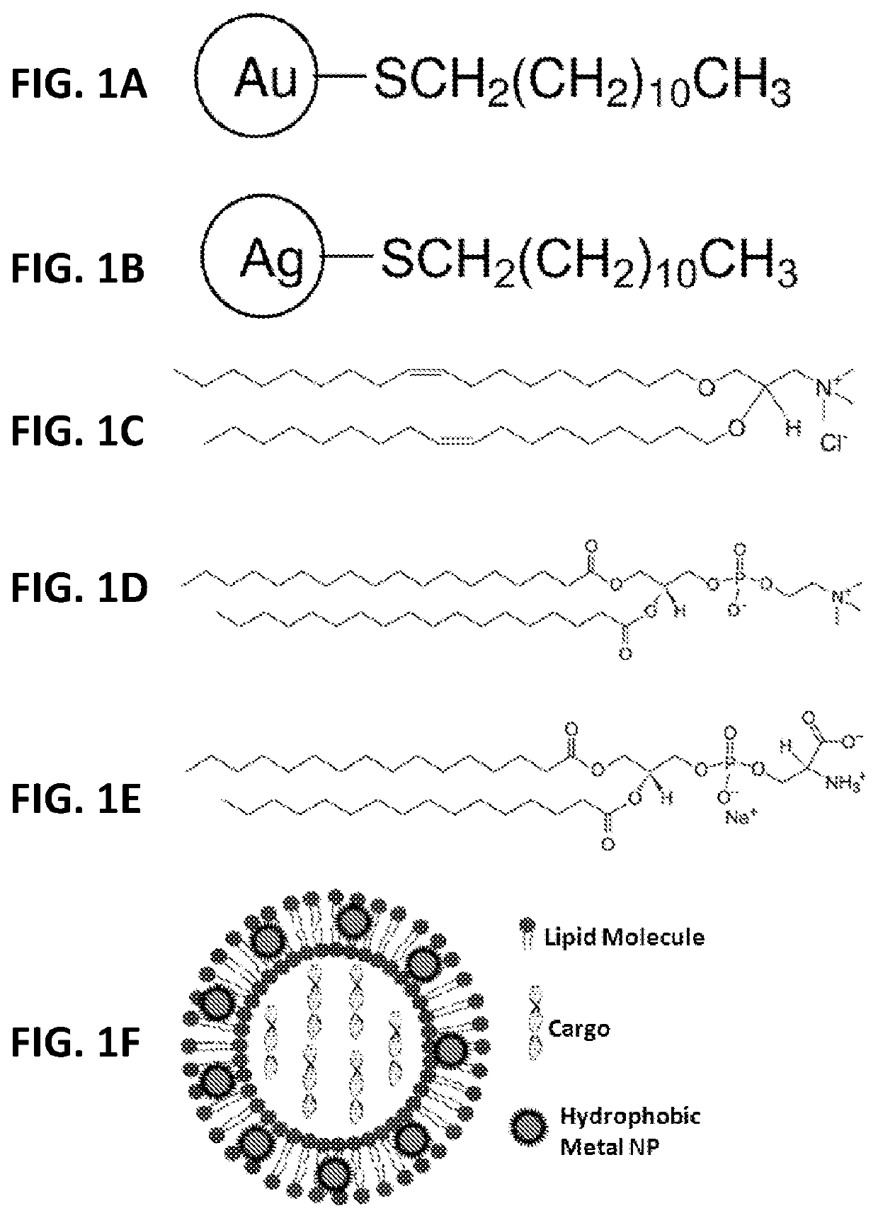
[0035]As shown in FIG. 1, numerous hydrophobic NPs, such as gold and silver NPs coated with various hydrophobic materials, such as dodecanthiol (commercially available from nanoComposix (San Diego, Calif., USA) with a diverse size range, including NPs with a mean diameter of 2, 4, and 5 nm, were incorporated into the bilayers of liposomes comprising: (a) a nucleic acid (e.g., pDNA or siRNA molecules) and perhaps other active agents encapsulated in the hydrophilic core; (b) a cationic lipid (e.g., DOTMA) comprising from about 50 mol % to about 100 mol % of the total lipid present in the liposome; and (c) a non-cationic lipid (e.g., DSPC) comprising from about 0.01 mol % to about 50 mol % of the total lipid present in the liposome. While the range of nanoparticles useful for practicing the present invention generally range from about 0.1 nm to about 20 nm in average diameter, a preferred range of average diameter is between about 0.1 nm to about 5 nm.

[0036]In one embodiment, liposomes...

example 2

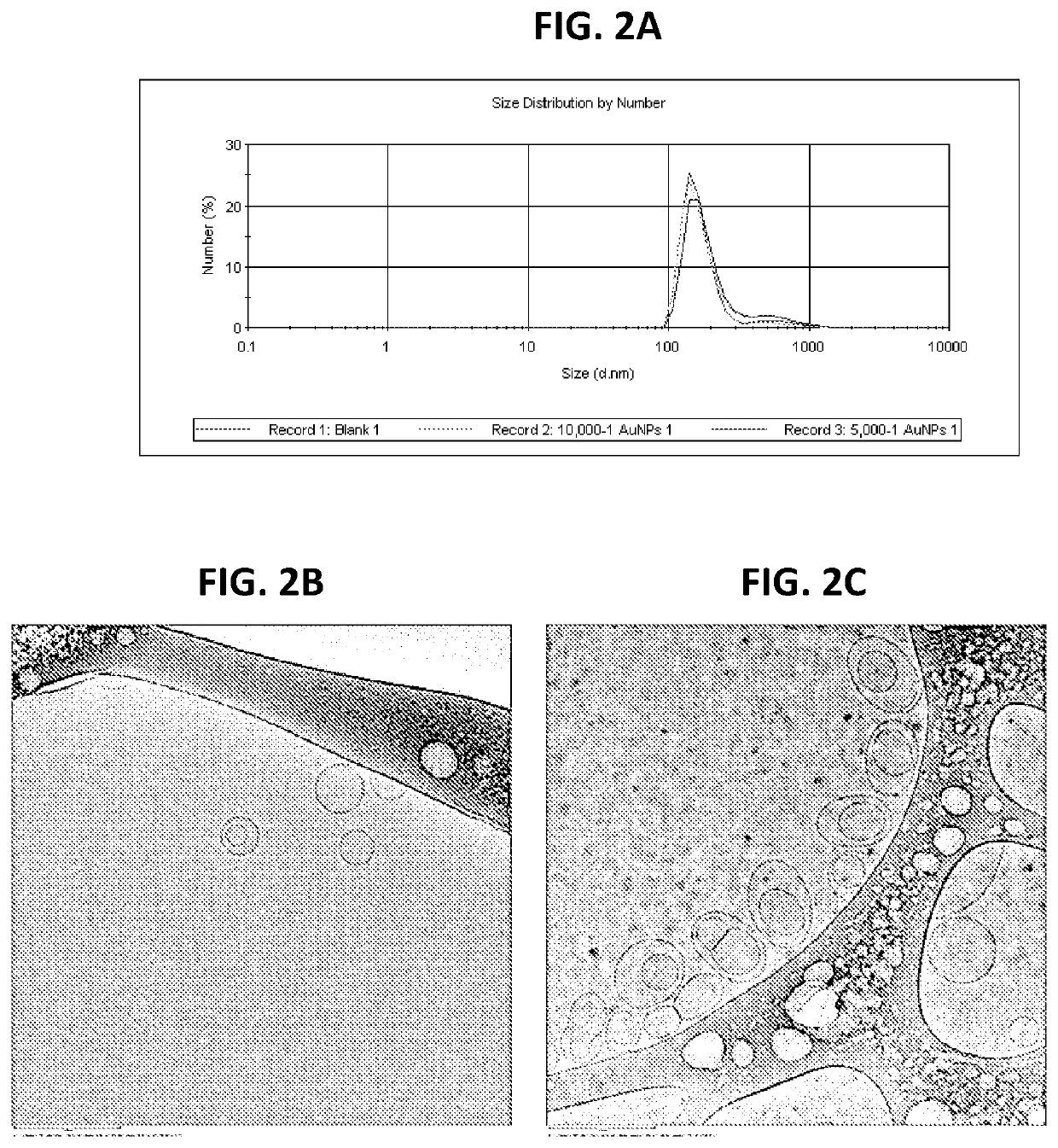
[0038]In another embodiment, MLVs were prepared by a thin film hydration method in order to study membrane fusogenicity enhancement induced by the inclusion of NPs in the membranes of these vesicles. Briefly, chloroform solutions of the cationic lipid DOTMA and the anionic lipid DPPS at an equimolar ratio with or without hydrophobic NPs at 10,000:1 or 5,000:1 (lipid molecules v. NPs) ratios were prepared in sterile glass vials. Thin uniform films were then prepared by rapidly evaporating the chloroform under vacuum for 2 hours at 25° C. in order to remove the trace solvent impurities. This film was then hydrated using an appropriate volume of citrate buffer (pH 4.5) in 10% D2O by vortexing for 30 seconds. The total lipid concentration in the resulting aqueous dispersion was 15 mM. The dispersion was then sonicated using a bath sonicator for 30 minutes. The morphology of the liposomes was assessed using cryo-TEM (JEM-2100F, Jeol USA Inc., MA, USA) and the phase transition was detecte...

example 3

[0039]In another embodiment, liposomes were prepared using a thin film hydration method. Briefly, a chloroform solution of the cationic lipid DOTMA and the non-ionic lipid DSPC at an equimolar ratio both with and without hydrophobic NPs at 10,000:1 (lipid molecules v. NPs) ratio was prepared in a sterile glass vial. A thin uniform film was then prepared by rapidly evaporating the chloroform under vacuum for 2 hours at 25° C. in order to remove the solvent. This film was then hydrated using an appropriate volume of phosphate buffered saline with / without comprising nucleic acid (pDNA) by vortexing for 30 seconds. The resulting dispersion was then sonicated using a bath sonicator for 30 minutes. The total lipid and pDNA concentrations in the liposomes were 2 mM and 40 ng / μl, respectively.


PUM

PropertyMeasurementUnit
phase transition temperatureaaaaaaaaaa
mol %aaaaaaaaaa
mol %aaaaaaaaaa

Abstract

The disclosure provides compositions and methods of use of formulations effective for the delivery of biologically active molecules and materials, including therapeutic and diagnostic agents, and combinations thereof, to cells, tissues, and living organisms. More particularly, the present invention is related to the incorporation of hydrophobic nanoparticles, such as hydrophobic metal nanoparticles, into the membranes of vesicular delivery vehicles. These hydrophobic nanoparticles enhance the endosomal membrane fusogenicity of the formulations through the promotion of inverted hexagonal phase formation in the lipid bilayers of the vesicular delivery vehicles. As a result, the vesicular delivery vehicles more readily fuse with the endosomal membranes, leading to enhanced endosomal escape of the vesicular delivery vehicle and its contents, thereby facilitating the delivery of the biologically active molecules and materials incorporated within the vesicular delivery vehicles to their sites of action.

Description

CROSS-REFERENCE TO RELATED APPLICATIONS[0001]This application claims priority to and the benefit of U.S. provisional patent application Ser. No. 62 / 479,753, filed Mar. 31, 2017, which application is incorporated herein by reference in its entirety.FIELD OF INVENTION[0002]The present invention relates to vesicular delivery vehicles that deliver therapeutic and diagnostic agents into living cells, more specifically, liposomes incorporated with nanoparticles for quicker and easier endosomal escape of the liposomes and their contents.BACKGROUND OF THE INVENTION[0003]Biological drugs, or biologics, such as oligonucleotides, proteins, peptides, enzymes, plasmid DNA (pDNA), small interfering RNA (siRNA), and antibodies, are effective and typically target-specific agents for the prevention, palliation, and treatment of disease. These biologically active compounds may have therapeutic targets in the cytoplasm, in the nucleus, or on the surface of cellular structures, membranes, and organelle...

Claims


Application Information

Patent Timeline
Patent Type & Authority Patents(United States)
IPC IPC(8): A61K9/127A61K47/02A61K47/20A61K47/18A61K31/7088C12N15/113A61K47/26
CPCA61K9/1271A61K31/7088A61K47/02A61K47/186A61K47/20A61K47/26C12N15/113C12N2310/14C12N2320/32
Inventor SARODE, ASHISHVERMA, RUCHIWORTHEN, DAVIDDENG, RUITANG
Owner UNIV OF RHODE ISLAND BOARD OF TRUSTEES
Why Eureka
Try Eureka