Method for neural current imaging

a neural current and imaging technology, applied in the field of neural current imaging, can solve the problems of limited spatial resolution, inability to identify sources with great accuracy, and cost-intensive shielding

Active Publication Date: 2003-05-15
MAGSTIM GRP INC
View PDF42 Cites 22 Cited by
  • Summary
  • Abstract
  • Description
  • Claims
  • Application Information

AI Technical Summary

Problems solved by technology

However, a disadvantage of EEG is that it has limited spatial resolution, i.e., it is unable to identify with great accuracy the spatial location of the sources of electrical activity within the brain.
Such shielding is costly to provide, and MEG cannot be used simultaneously with other techniques producing electromagnetic radiations.
Because the energy states for the spin angular momenta are quantized, these interactions must permit precise amounts of energy loss or relaxation cannot occur, and due to the randomness of the interactions, it results that a substantial time is required for relaxation.
Where the fluctuations occur at rates that are either to large or too small compared to the Larmor frequency, energy dissipation is inefficient and therefore slow, resulting in long decay times T.sub.1 and T.sub.2.
Even so, the spectra do not contain any spatial information so that an image cannot yet be formed.
A disadvantage of MRI is that the time required for spin relaxation, even the relatively short time required for relaxation of protons in lipids, limits the temporal resolution of MRI.
However, MRNCI has not yet been shown to be able to detect currents as small as neural cu

Method used


Image

  • Method for neural current imaging
  • Method for neural current imaging
  • Method for neural current imaging

Examples


Embodiment Construction

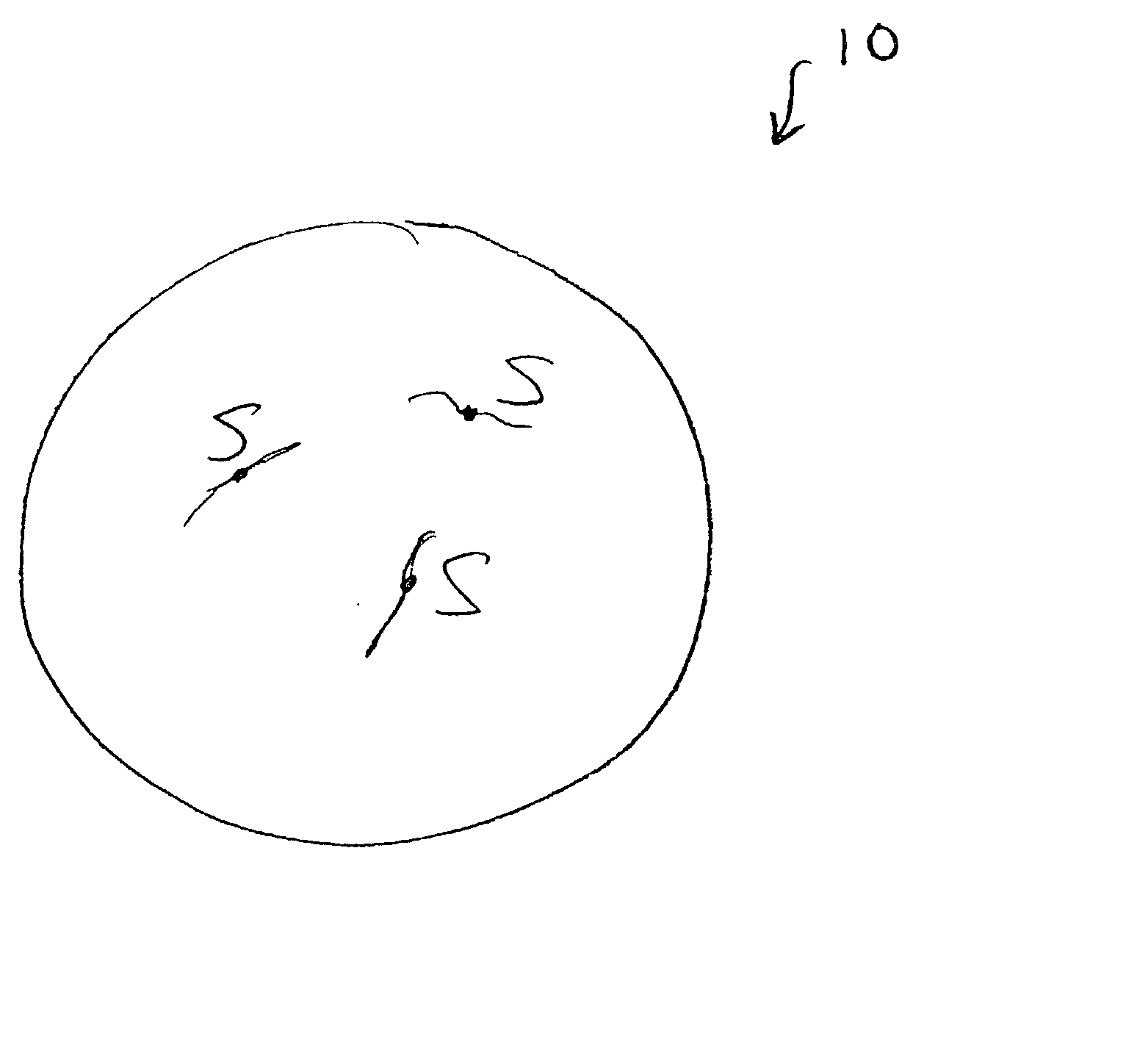
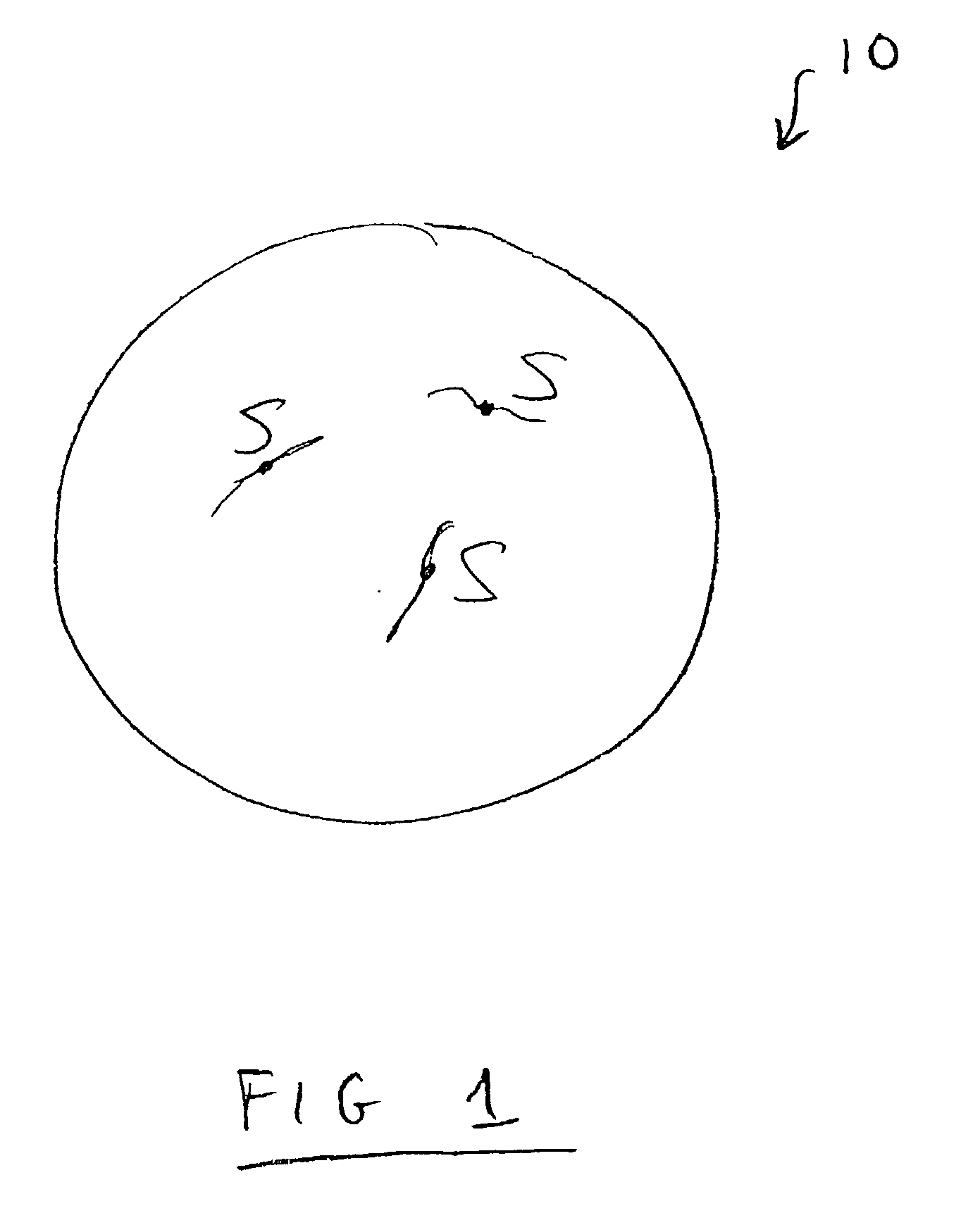
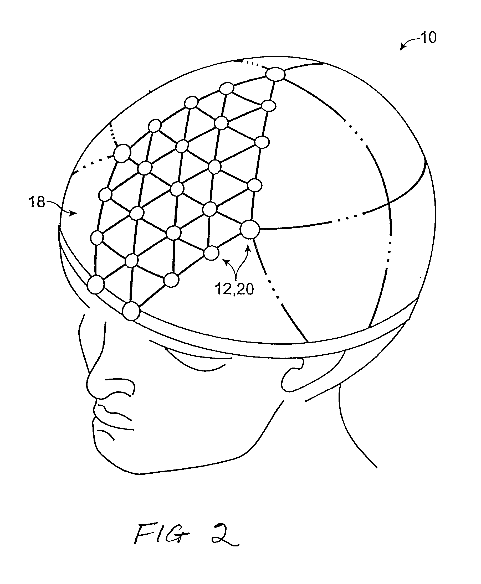
[0037] A preferred method for imaging neural currents according to the present invention is described below, along with the preferred application of determining the spatial and temporal parameters or coordinates of neural currents in the brain. However, it should be understood that methods according to the invention may be used generally to improve the clarity or precision of any data and are therefore not limited to neural current imaging. Moreover, methods according to the present invention may be used for imaging any electrophysiological activity in which current flows, such as heart electrical activity.

[0038] Referring to FIG. 1, a portion 10 of a body, such as a human head, is shown with sources "S" inside that are representative of neural activity. Particularly, the sources "S" represent nerve cells that carry neural currents from one interior location in the head to another. The neural activity can result from a voluntary or involuntary motor function, or be a result of thoug...


PUM


Abstract

A method for neural current imaging. Electromagnetic fields produced unknown distribution of unknown sources in the body is sensed at a plurality of remote locations. The inverse problem is solved to produce a first fuzzy "image" of the sources. In addition, a standard imaging method, such as MRI, is used to independently image the body, to obtain a second fuzzy image of the sources. Additional independently obtained fuzzy images may also be provided. All or a selected subset of the images are pooled as components to form an enhanced image with greater resolution or clarity than the component images.

Description

[0001] This is a continuation-in-part of U.S. Ser. No. 10 / 001,541, now pending.[0002] The present invention relates to a method for imaging neural current, for detecting and locating neural activity in the body, and more particularly, in the brain.BACKGROUND OF INVENTION[0003] It is well known that electrical currents are associated with brain activity. These electrical currents are produced by neurons in the brain and are referred to as "neural currents." Information about neural currents provides a means for understanding brain function. For example, the performance of a specific motor function will involve particular areas of the brain, and the dynamic behavior of the neural currents associated with the motor function provides information about the sequence in which different areas of the brain are involved.[0004] Electroencephalography ("EEG") is a technique that may be used for measuring neural currents. In EEG, electrodes are placed at various locations on the scalp and the di...

Claims


Application Information

Patent Timeline
IPC IPC(8): A61B5/04A61B5/053
CPCA61B5/04A61B5/7264A61B5/0536A61B5/4064A61B5/24
Inventor TUCKER, DON M.
Owner MAGSTIM GRP INC
Why Eureka
Try Eureka