
Radiation image radiographic apparatus

a radiographing apparatus and radiation image technology, applied in the direction of instruments, tubes with screens, patient positioning for diagnostics, etc., can solve the problems of insufficient contrast of radiographed images, inability of radiation image radiographing apparatus to radiograph an image clear, and countless changes in the magnifying rate of images

Inactive Publication Date: 2004-06-10
KONICA MINOLTA MEDICAL & GRAPHICS INC
View PDF17 Cites 22 Cited by
  • Summary
  • Abstract
  • Description
  • Claims
  • Application Information

AI Technical Summary

Benefits of technology

[0013] According to the radiation image radiographing apparatus of the first aspect of the present invention, since the radiation image radiographing apparatus comprises a plurality of radiation image information detecting members, an operator only needs to choose any one among the plurality of radiation image information detecting members for performing radiography. Therefore, the operator does not have to make a fine adjustment on a magnifying rate or the like, and it is possible to easily repeat the radiography of a predetermined magnifying rate. In other words, in both the cases of radiographing a normal absorption contrast image and radiographing a phase contrast image, since the operator only needs to choose any one of the plurality of radiation image information detecting members attached to each of the plurality of supporting platforms for switching a radiography mode, it is possible to easily switch a radiography mode. Further, since it is also possible to switch a mode of magnifying radiography by attaching a radiation image information detecting member to a predetermined supporting platform or the like, it is possible to simplify a structure of the apparatus, and thereby it is possible to improve user-friendliness and reduce cost on the apparatus.
[0050] According to the apparatus, since at least two of the plurality of supporting platforms are located in the space opposite to the radiation source with respect to the subject platform and therefore they could interfere radiography, by evacuating the at least two supporting platforms from the space, it is possible to easily perform radiography.

Problems solved by technology

However, since the subject is radiographed at substantially full scale with this method, contrast of the radiographed image is insufficient.
Thereby, the radiation image radiographing apparatus is not capable of radiographing an image clear enough to provide fine structure of the specific region of a body for interpretation.
However, with the method in the earlier art, since the supporting platform is capable of moving its location arbitrarily, a magnifying rate of the image also changes countlessly.
Consequently, when it is necessary to obtain images with the same magnifying rate, even though calibration of the magnifying rate is provided, it is not easy to make a fine adjustment.
Further, since it is also necessary to have a location recognition device as a means to recognize a location for obtaining information of distance between a radiation source and the subject platform (with which the subject is in contact) and the distance between the subject platform and the radiation image information detecting member, control radiation dose based on the information and the like, the control becomes more complicated.
Furthermore, since it is necessary to have such a complicated control means as mentioned above, it causes an increase on cost of the apparatus.

Method used


Image

  • Radiation image radiographic apparatus
  • Radiation image radiographic apparatus
  • Radiation image radiographic apparatus

Examples


first embodiment

[0065] [First Embodiment]

[0066] Hereinafter, an embodiment of the invention will be explained. However, the invention is not limited to the embodiment explained hereafter. Further, there are descriptions where meanings of words are explained hereinafter, but the descriptions are only to explain the meanings of the words, and the meanings of the words are not limited to the descriptions.

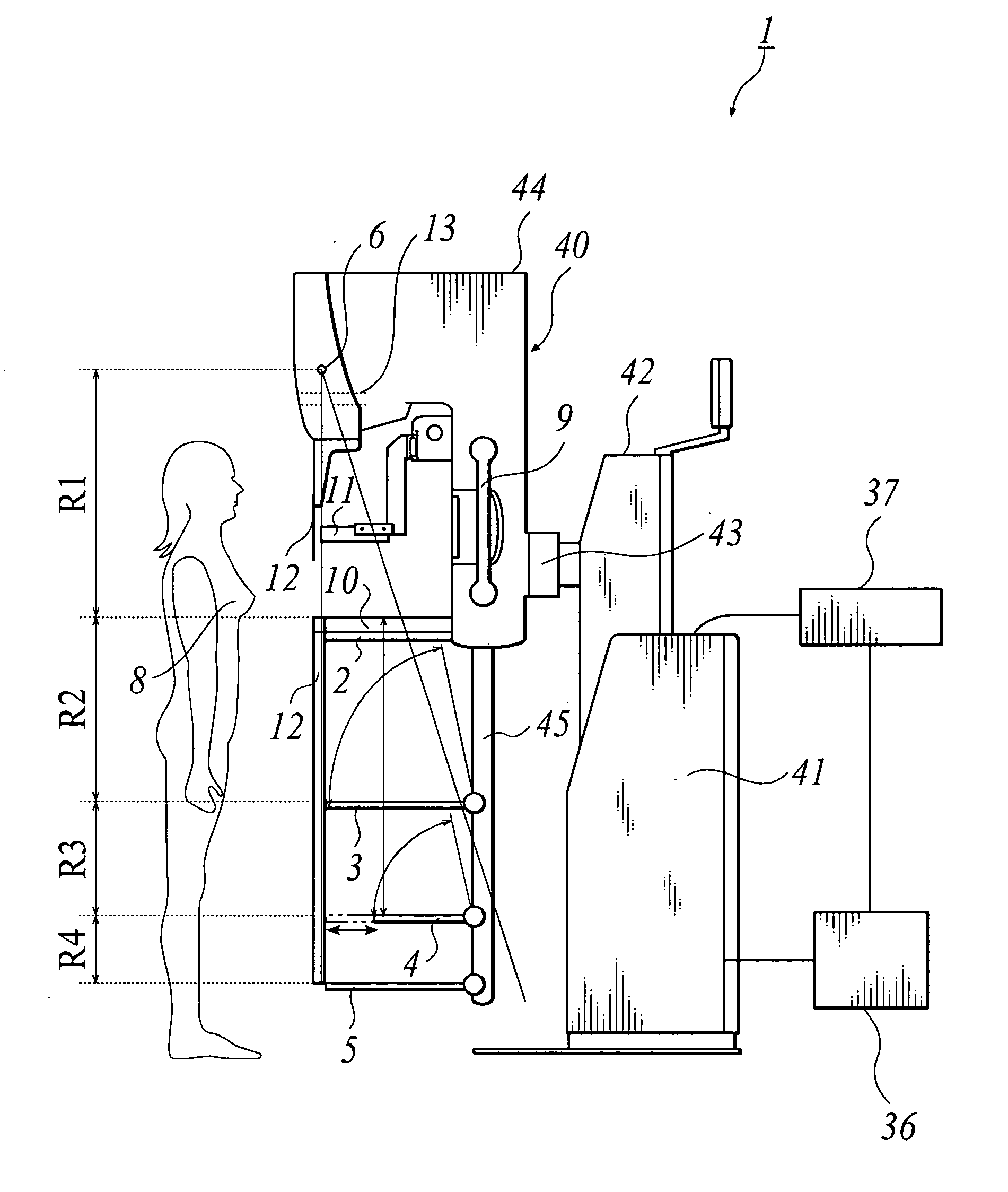
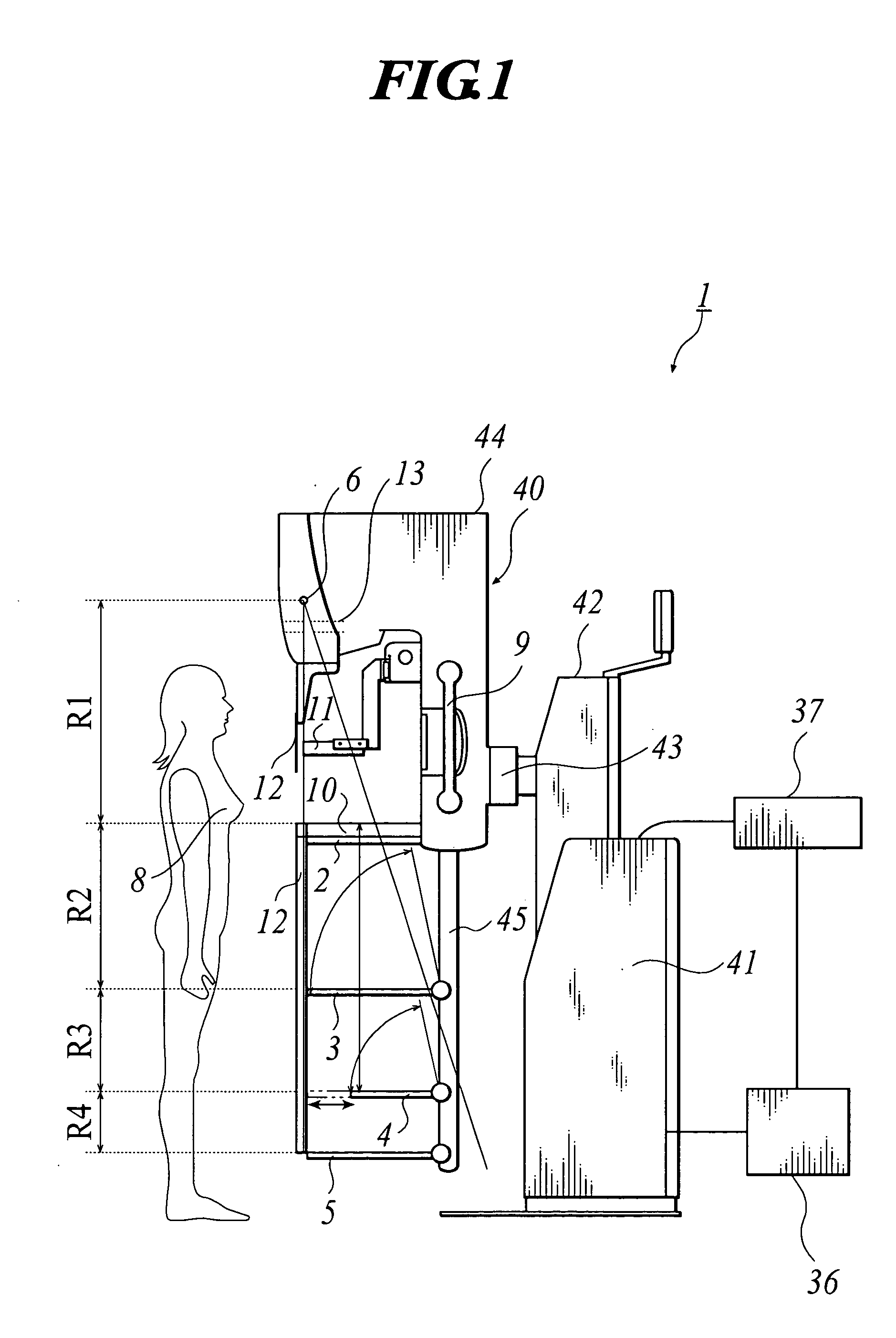
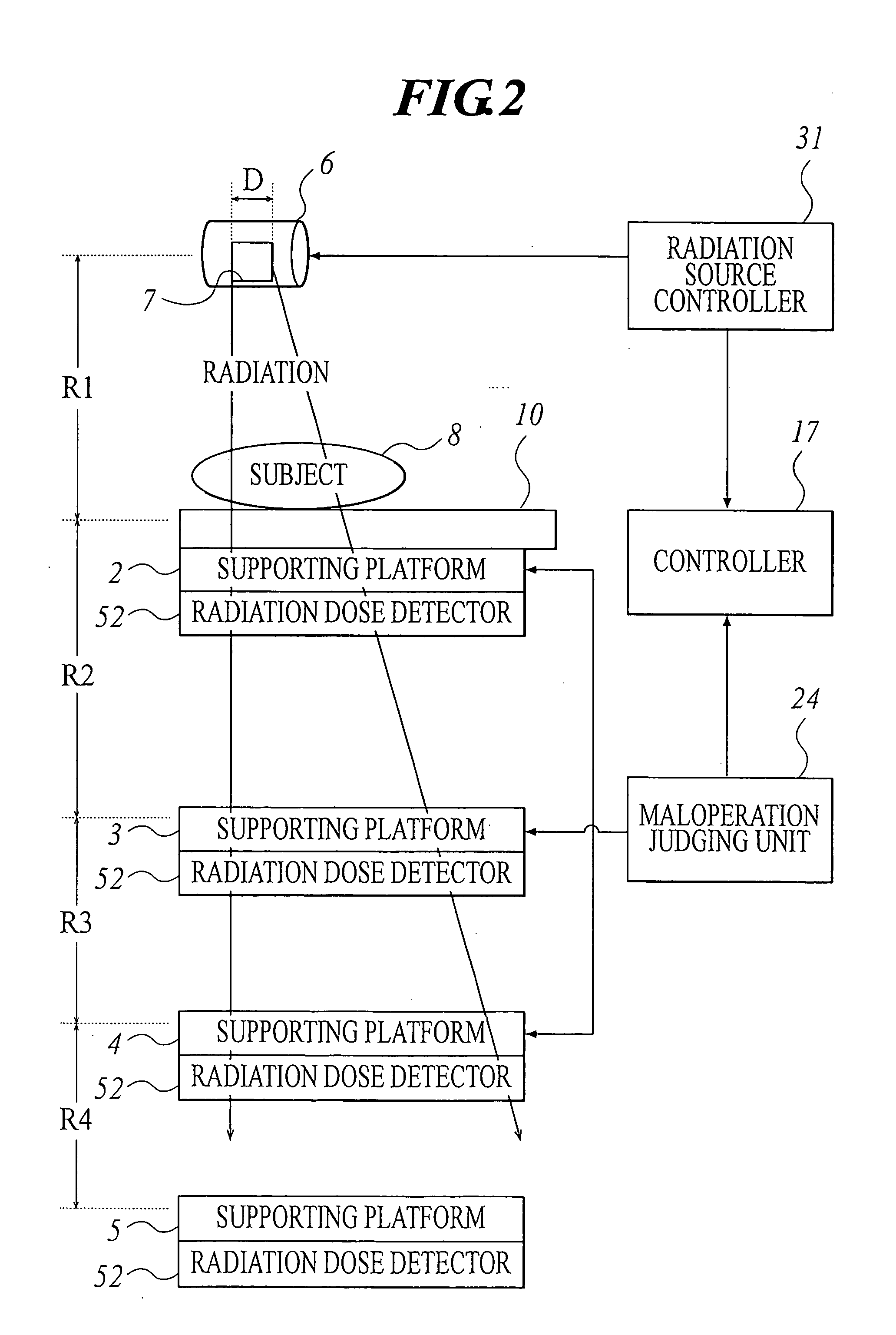
[0067] FIG. 1 is a side view showing a radiation image radiographing apparatus 1 in a first embodiment applied to the present invention. FIG. 2 is a pattern diagram showing a whole structure of the radiation image radiographing apparatus 1.

[0068] As shown in FIG. 1, the radiation image radiographing apparatus 1 comprises an apparatus body 40, a controller 17 (shown in FIG. 4), a radiation operation panel 37 having keys for selecting a radiography mode, a power supply 36 as a power source of the whole radiation image radiographing apparatus 1, supporting platforms 2, 3, 4 and 5 for supporting radiation...

second embodiment

[0137] [Second Embodiment]

[0138] Hereinafter, with reference to figures, a second embodiment applied to the present invention will be explained.

[0139] FIG. 5 is a side view showing a radiation image radiographing apparatus 101 in the second embodiment applied to the present invention. As shown in FIG. 5, in the radiation image radiographing apparatus 101, to each part, a code having lower two digits equal to the corresponding part in the radiation image radiographing apparatus 1 in the first embodiment is distributed.

[0140] As shown in FIG. 5, the radiation image radiographing apparatus 101, as well as the radiation image radiographing apparatus 1, comprises an apparatus body 140, a radiation operation panel 137, a power supply 136, supporting platforms 103 and 104, and a supporting member 145.

[0141] The apparatus body 140 has a first supporting base 141, a second supporting base 142, a spindle 143 and a radiography unit 144. To the first supporting base 141, an operation device 150...


PUM


Abstract

A user-friendly, inexpensive radiation image radiographing apparatus capable of easily switching between absorption contrast image radiography and phase contrast image radiography. The radiation image radiographing apparatus has: a radiation source; a subject platform for supporting a subject so as to face the subject to the radiation source; and a plurality of supporting platforms for supporting a radiation image information detecting member at a side opposite to the radiation source with respect to the subject platform, the radiation image information detecting member for detecting radiation image information based on radiation transmitted through the subject, wherein distances between the plurality of supporting platforms and the radiation source are different from each other.

Description

[0001] 1. Field of the Invention[0002] The present invention relates to a radiation image radiographing apparatus, in particular, a radiation image radiographing apparatus capable of radiographing a phase contrast image.[0003] 2. Related Art[0004] Generally, a radiation image radiographing apparatus using a mechanism of transmitting radiation through a substance is widely used for a medical image diagnosis, a non-destructive inspection and the like. In particular, in a radiation image radiographing apparatus for mammography, which is used for radiographing a specific region of body, radiography of a subject fixed on a subject platform which is combined with a radiation image information detecting member has been performed. However, since the subject is radiographed at substantially full scale with this method, contrast of the radiographed image is insufficient. Thereby, the radiation image radiographing apparatus is not capable of radiographing an image clear enough to provide fine ...

Claims


Application Information

Patent Timeline
Patent Type & Authority Applications(United States)
IPC IPC(8): A61B6/00A61B6/04
CPCA61B6/0414A61B6/107A61B6/4216A61B6/4233A61B6/5235A61B6/4494A61B6/484A61B6/502A61B6/4283
Inventor AMITANI, KOUJIMITSUMOTO, TAKAHIRO
Owner KONICA MINOLTA MEDICAL & GRAPHICS INC
Learn More