

Method and system for classifying slides using scatter plot distribution
a scatter plot and distribution method technology, applied in image data processing, instruments, image enhancement, etc., can solve the problems of limiting the traditional pap smear method, pap smear tests, and other tests, so as to reduce the number of slides
- Summary
- Abstract
- Description
- Claims
- Application Information
AI Technical Summary
Benefits of technology
Problems solved by technology
Method used
Image

Examples
Embodiment Construction
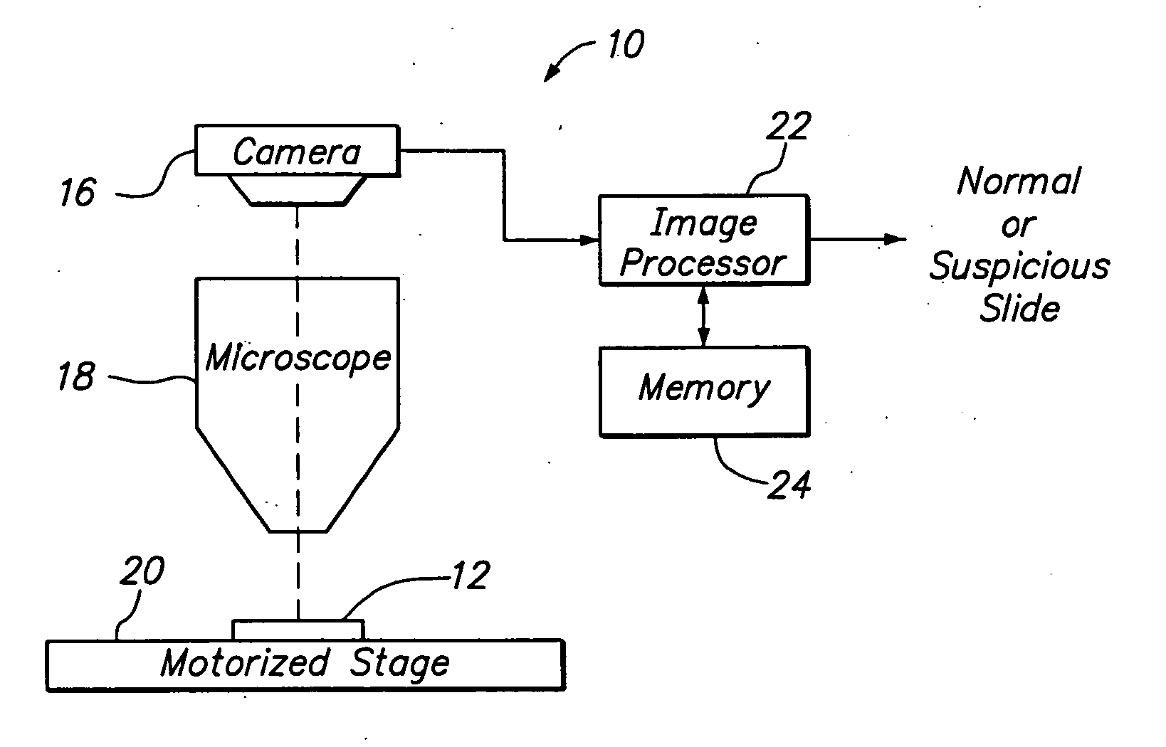
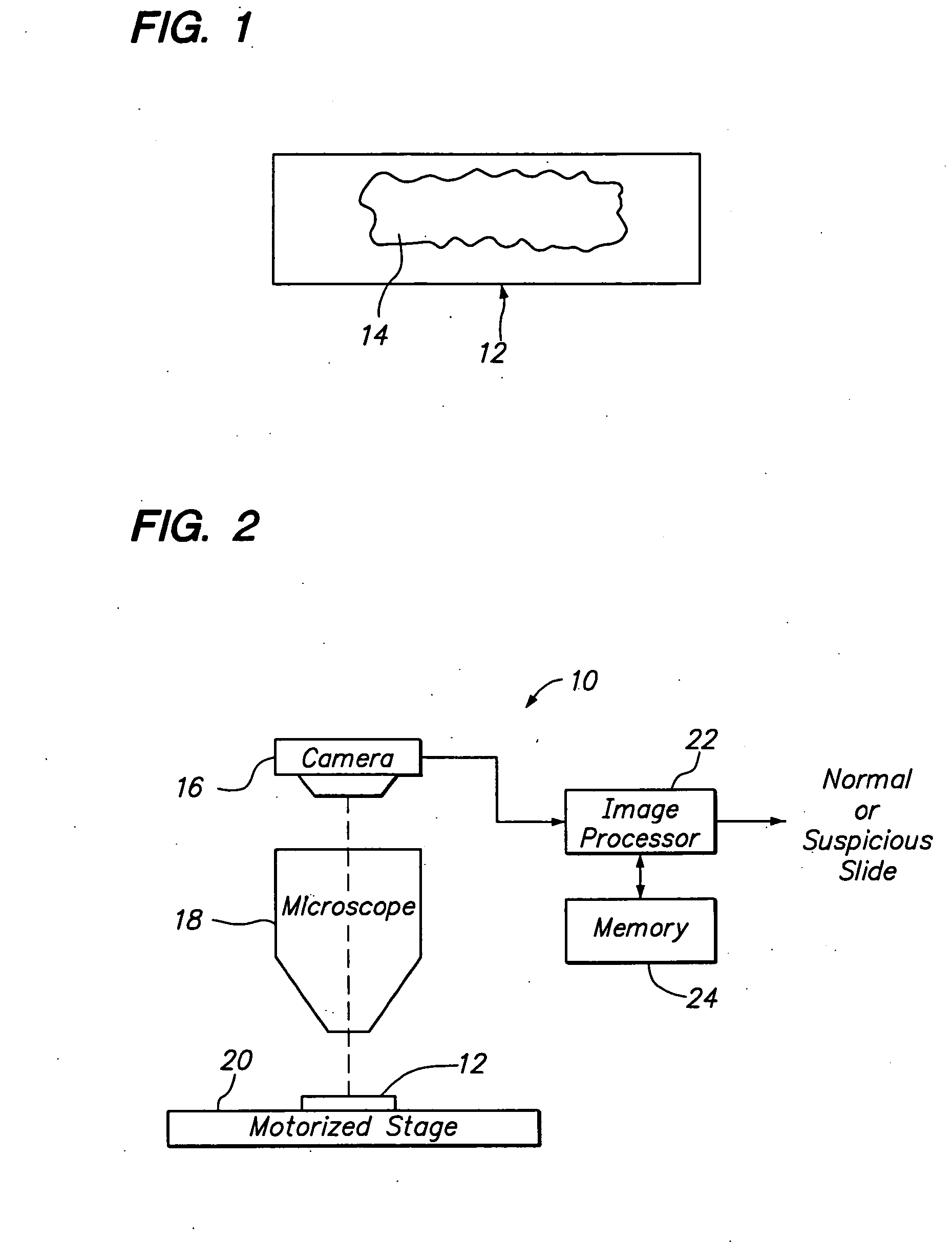
[0025] Referring to FIG. 2, a biological screening system 10 constructed in accordance with one preferred embodiment of the present invention is described. The system 10 is configured to process a series of microscope slides 12 in order to classify a biological specimen 14 (best shown in FIG. 1) as either "normal," which would not require review by a cytotechnologist, or "suspicious," which would require review by the cytotechnologist.
[0026] Although the system 10 can be used to classify any biological specimen as normal or suspicious, the system 10 lends itself particularly well to the presentation of cytological cervical or vaginal material, such as that typically found on a Pap smear slide. In this case, the cells may reflect abnormalities (e.g., cytolysis, atrophy, infection, damage), malignancy or premalignancy, such as Low Grade Squamous Intraepithelial Lesions (LGSIL) or High Grade Squamous Intraepithelial Lesions (HGSIL), as well as all other cytologic categories as defined ...
PUM
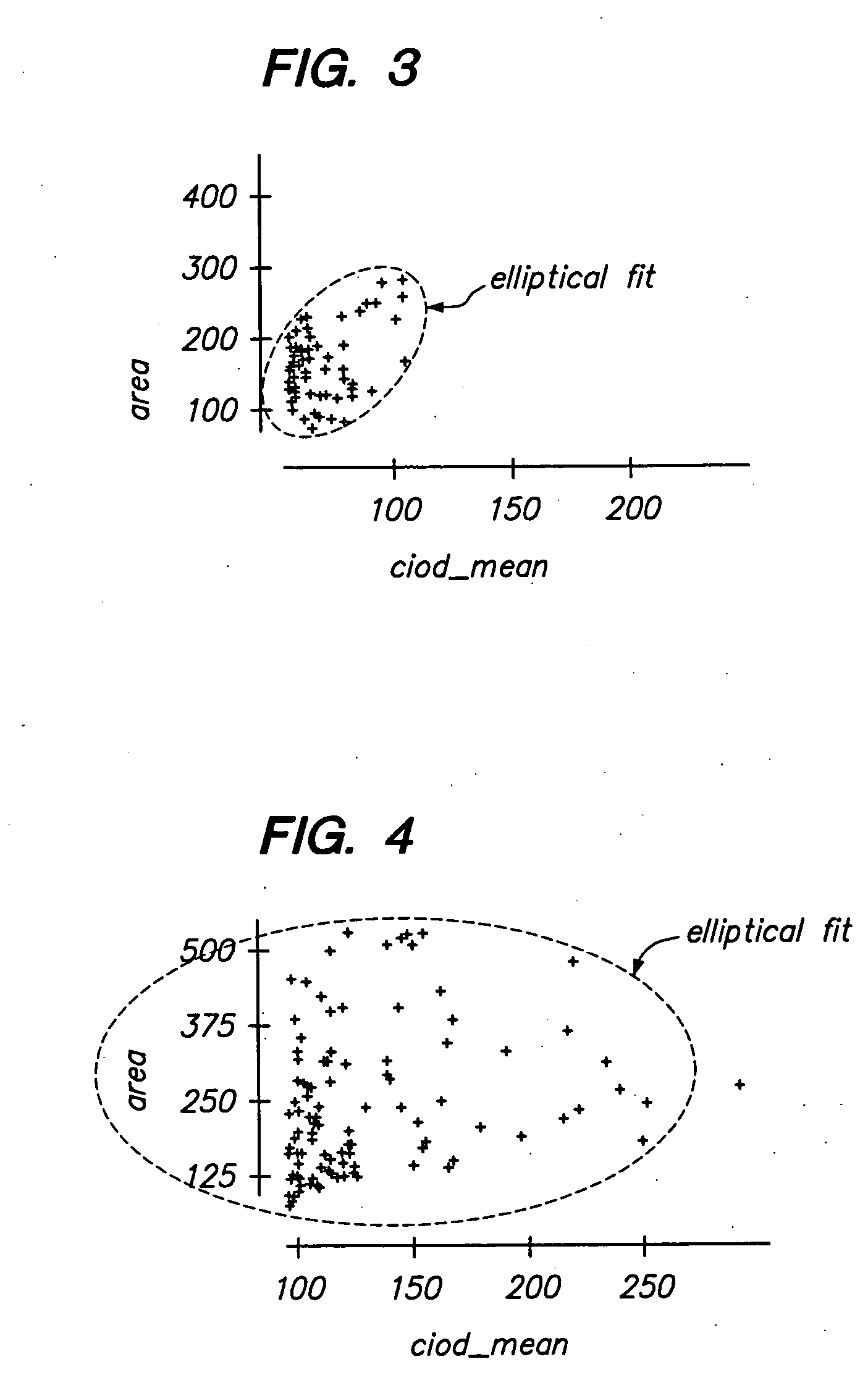
| Property | Measurement | Unit |
|---|---|---|
| diameter | aaaaa | aaaaa |
| nuclear area | aaaaa | aaaaa |
| nuclear integrated optical density | aaaaa | aaaaa |
Abstract
Description
Claims
Application Information