
Method and apparatus for measuring a retinal sublayer characteristic

a technology of retinal sublayer and characteristic, applied in the field of eye diagnostic evaluation apparatus, can solve the problems of gradual loss of vision, inability to restore lost neural tissue, and inability to detect changes in visual field

Active Publication Date: 2005-01-27
THE CLEVELAND CLINIC FOUND
View PDF5 Cites 228 Cited by
  • Summary
  • Abstract
  • Description
  • Claims
  • Application Information

AI Technical Summary

Benefits of technology

In accordance with another aspect of the present invention a computer program product, recorded in a computer readable medium and operative in a data processing system, is provided for measuring the thickness of a retinal layer within of an eye. A scanner control component directs an associated scanner to conduct a plurality of axial scans of a desired area of the retina of the eye and measure a set of reflection intensity values for each axial scan. A fine boundary search component performs a progressive refinement boundary detection algorithm using the measured sets of reflection values to determine boundary locations of the retinal layer. A layer thickness mapping component maps the thickness of the retinal layer across the desired area of the retina in response to the determined boundary locations of the retinal layer.
In accordance with still an...

Problems solved by technology

Glaucoma is a leading cause of blindness in the US, characterized by a gradual loss of vision due to damage to optic nerve tissue.
Glaucoma usually responds well to treatment, but treatment cannot restore lost neural tissue.
However, visual field changes may not be detectable until significant damage has been done to the RNFL.
In some cases, up to fifty percent of nerve fibers can be damaged before a visual field defect can be detected by perimetry.

Method used


Image

  • Method and apparatus for measuring a retinal sublayer characteristic
  • Method and apparatus for measuring a retinal sublayer characteristic
  • Method and apparatus for measuring a retinal sublayer characteristic

Examples


Embodiment Construction

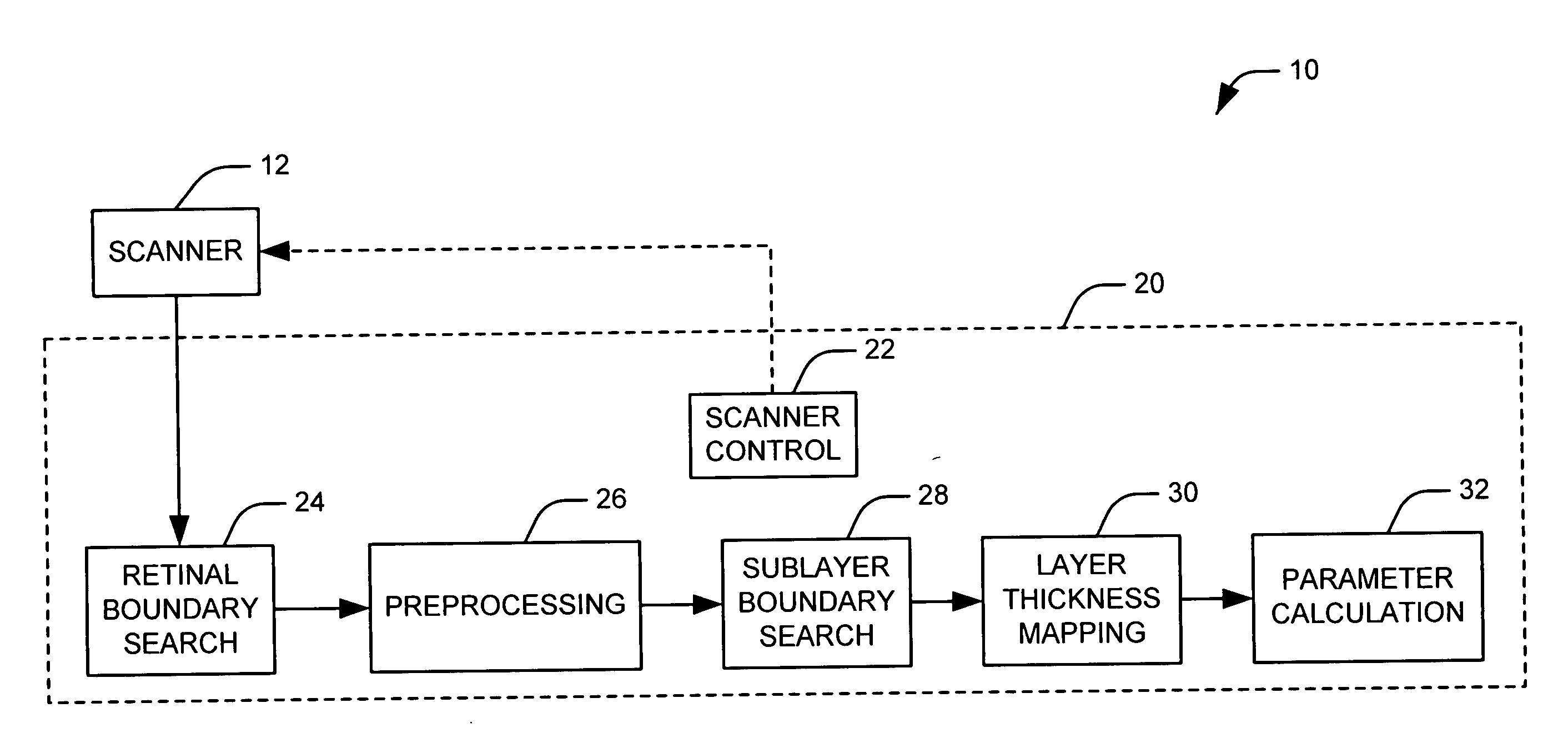
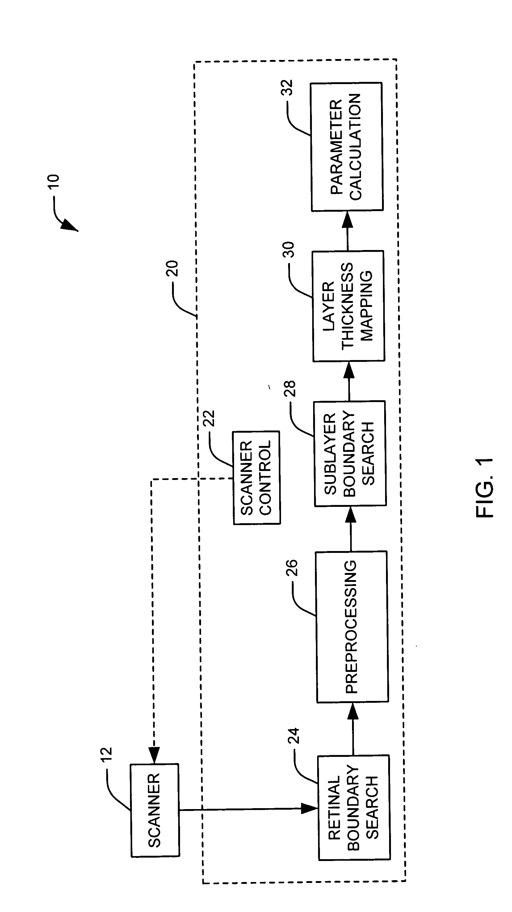
The present invention relates to an apparatus and method for diagnostic evaluation of the eye and, in particular, is directed to a method and apparatus for measuring a retinal sublayer characteristic. As representative of the present invention, FIG. 1 illustrates an assembly 10 for measuring one or more characteristics of a sublayer of a retina. The assembly includes a scanner 12 operative to perform axial scanning of a human or animal eye. The assembly further includes a control module 20 for the scanner 12 in accordance with a first embodiment of the present invention. It will be appreciated that the control module 20 and its various components can be implemented either as dedicated hardware circuitry appropriate for a described function or as computer software, recorded in a computer readable medium and operative to perform the described function when executed by a data processing system. For example, the control module can comprise a software module executed on a general purpos...


PUM


Abstract

Methods and systems are provided for measuring a retinal sublayer characteristic of an eye. A plurality of axial scans are performed over an area of the retina of the eye. Reflections are measured during the axial scans to determine a plurality of sets of reflection intensity values. A given set of reflection intensity values is associated with one of the plurality of axial scans. A progressive refinement boundary detection algorithm is performed using the plurality of sets of reflection intensity values to determine at least one boundary location associated with the retinal sublayer for each of the plurality of sets of reflection intensity values. The retinal sublayer characteristic is determined in response to the determined boundary locations.

Description

FIELD OF THE INVENTION The present invention relates to an apparatus and method for diagnostic evaluation of the eye and, in particular, is directed to a method and apparatus for measuring a retinal sublayer characteristic. BACKGROUND OF THE INVENTION Glaucoma is a leading cause of blindness in the US, characterized by a gradual loss of vision due to damage to optic nerve tissue. The ganglion cell layer (GCL) of the retina contains the cell bodies of the retinal nerve fibers, which travel through the retinal nerve fiber layer (RNFL) and coalesce at the optic nerve head (ONH) to form the optic nerve. One hallmark of glaucoma is the progressive loss of neural tissue in all these structures and consequent visual loss. Glaucoma usually responds well to treatment, but treatment cannot restore lost neural tissue. It is thus important to diagnose the disease quickly to prevent irreversible visual loss. Glaucoma diagnostic methods can generally be classified as functional or structural. ...

Claims


Application Information

Patent Timeline
IPC IPC(8): A61B3/10A61B3/12
CPCA61B3/102
Inventor HUANG, DAVIDTAN, OULI, YAN
Owner THE CLEVELAND CLINIC FOUND
Learn More