Method and device for detecting tissue boundaries by use of ultrasonic images

An ultrasound image and boundary technology, applied in image enhancement, image analysis, image data processing and other directions, can solve the problems of not meeting real-time performance, and the results of dual-region gray distribution functional cannot be obtained, and achieves simple detection and low dependence. , the effect of improving the calculation speed

Active Publication Date: 2009-09-09
SHENZHEN MINDRAY BIO MEDICAL ELECTRONICS CO LTD +1
View PDF0 Cites 51 Cited by
  • Summary
  • Abstract
  • Description
  • Claims
  • Application Information

AI Technical Summary

Problems solved by technology

Although this method can remove the influence of noise well, it cannot meet the real-time requirements, and the echocardiographic image cannot simply be regarded as an image containing only the foreground and background, that is, the image of the blood region of the left vent

Method used


Image

  • Method and device for detecting tissue boundaries by use of ultrasonic images
  • Method and device for detecting tissue boundaries by use of ultrasonic images
  • Method and device for detecting tissue boundaries by use of ultrasonic images

Examples


Embodiment Construction

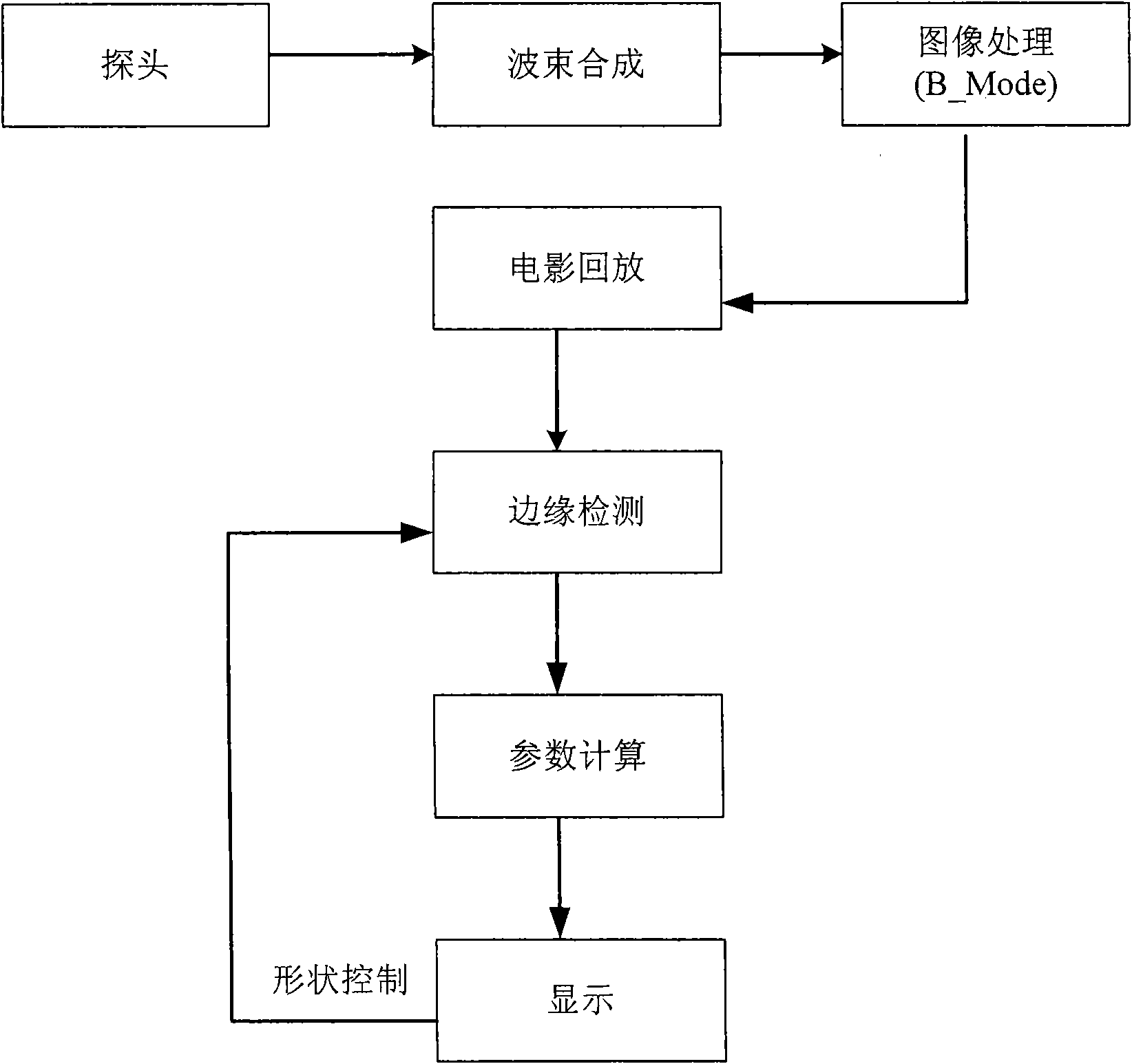
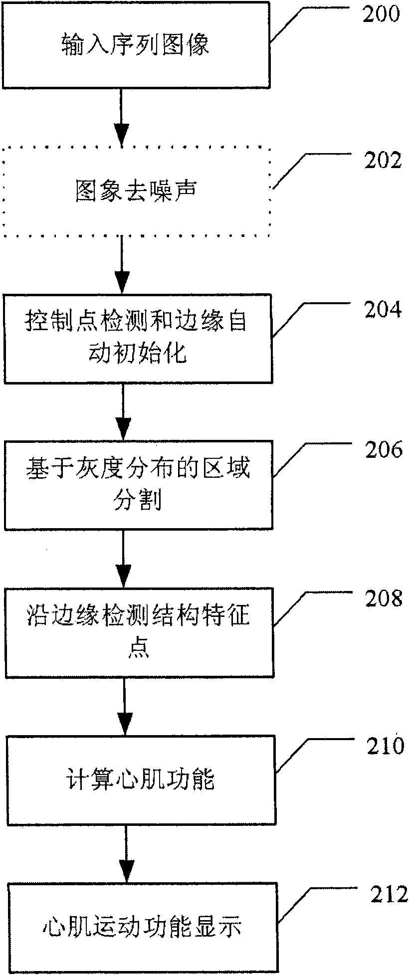
[0052] Taking the detection of the endocardial border as an example, the method and device according to this embodiment will be described in detail below.

[0053] like figure 1 Shown is a block diagram of a typical endocardial detection system. The ultrasonic probe emits ultrasonic waves to the corresponding examination parts of the human body (such as the heart). The imaging scan can be triggered and controlled by ECG. The received echo signal is processed by pre-amplification, ADC conversion, beam forming and other links, and then sent to the image processing module. After non-Doppler signal processing, the gray scale image of the anatomical structure of the human tissue (such as the heart) is obtained and stored in the movie playback data storage unit. The automatic boundary detection module reads the image data, performs boundary detection and calculation, and the parameter calculation module calculates the parameters of various functions (such as heart function) accordi...


PUM


Abstract

The invention discloses a method and a device for detecting tissue boundaries by use of ultrasonic images. The method comprises a search step and a detection step, wherein the search step is used for searching an ultrasonic image of a detection target for an initial center point of a target region and determining an initial region boundary curve according to the initial center point; the detection step takes the initial center point as a pole, starts searching for the position of a target region boundary along a polar line passing the pole from an initial region boundary, and calculates the energy functional of gray-scale distribution in different positions, wherein a position corresponding to the minimum value of the energy functional of gray-scale distribution is the boundary of the target region. As the invention first detects the center point and then rapidly detects the boundaries along the polar line, the invention is simpler and rapider in detection. Region segmentation functional adopted in the invention can effectively avoid the influence of ultrasonic image noise and effectively detects weak boundaries. In addition, grey scale correction along the polar line can eliminate the influence of non-uniform fields of image gray-scale distribution.

Description

technical field [0001] The invention relates to an ultrasonic image analysis technology, in particular to a method and device for detecting tissue boundaries using an ultrasonic image. Background technique [0002] Ultrasonography has become an important and non-invasive detection method for displaying the structure and motor function of human organs, and further quantitative analysis of the structure and function of these organs first needs to determine the boundary information of these organs. For example, the quantitative analysis of cardiac function requires the measurement of shape parameters such as the area of ​​the cardiac chamber section and its changes during the cardiac cycle, which requires a clear endocardial boundary. At the same time, border delineation can also be used to track the lesion. In addition, the 3D reconstruction of human organ images also needs to outline the boundaries of the organs as a basis. [0003] As the most commonly used field of human ...

Claims


Application Information

Patent Timeline
IPC IPC(8): G06T7/60G06T5/00A61B8/00
CPCG06T7/0081G06T2207/10132G06T7/0089G06T2207/30048G06T7/149G06T7/11
Inventor 李鑫丛龙飞张羽
Owner SHENZHEN MINDRAY BIO MEDICAL ELECTRONICS CO LTD
Why Eureka
Try Eureka