Suprabasal breast cell with stem cell properties

a breast cell and stem cell technology, applied in the field of breast cell with stem cell properties, can solve the problems of not being able to characterise the putative stem cell to show the full potential of generating tdlu, and achieve the effect of generating tdlu and demonstrating the full potential of tdlu

Inactive Publication Date: 2005-11-03
UNIVERSITY OF COPENHAGEN +1
View PDF1 Cites 11 Cited by
  • Summary
  • Abstract
  • Description
  • Claims
  • Application Information

AI Technical Summary

Benefits of technology

[0025] In a further aspect of the invention the cells isolated by the method disclosed in the present application are expanded and provides a method of transplanting a vertebrate host with said cells, comprising the step of introducing the cell into the vertebrate host. In yet a further development of this aspect said cells provide a method of in vivo administration of a protein or gene of interest to an individual in need thereof, comprising the step of transfecting the cell-population with a vector comprising DNA or RNA which expresses the protein or gene of interest and introducing the transfected cell into said individual. In yet a further development of the invention said cells provide a method of tissue repair or transplantation in mammals, comprising administering to a mammal a therapeutically effective amount of cells or tissues derived therefrom.

Problems solved by technology

However, further characterisation of the putative stem cells to show the full potential of generating TDLU have not been pursued due to a limited growth potential in primary culture.

Method used


Image

  • Suprabasal breast cell with stem cell properties
  • Suprabasal breast cell with stem cell properties
  • Suprabasal breast cell with stem cell properties

Examples


example 1

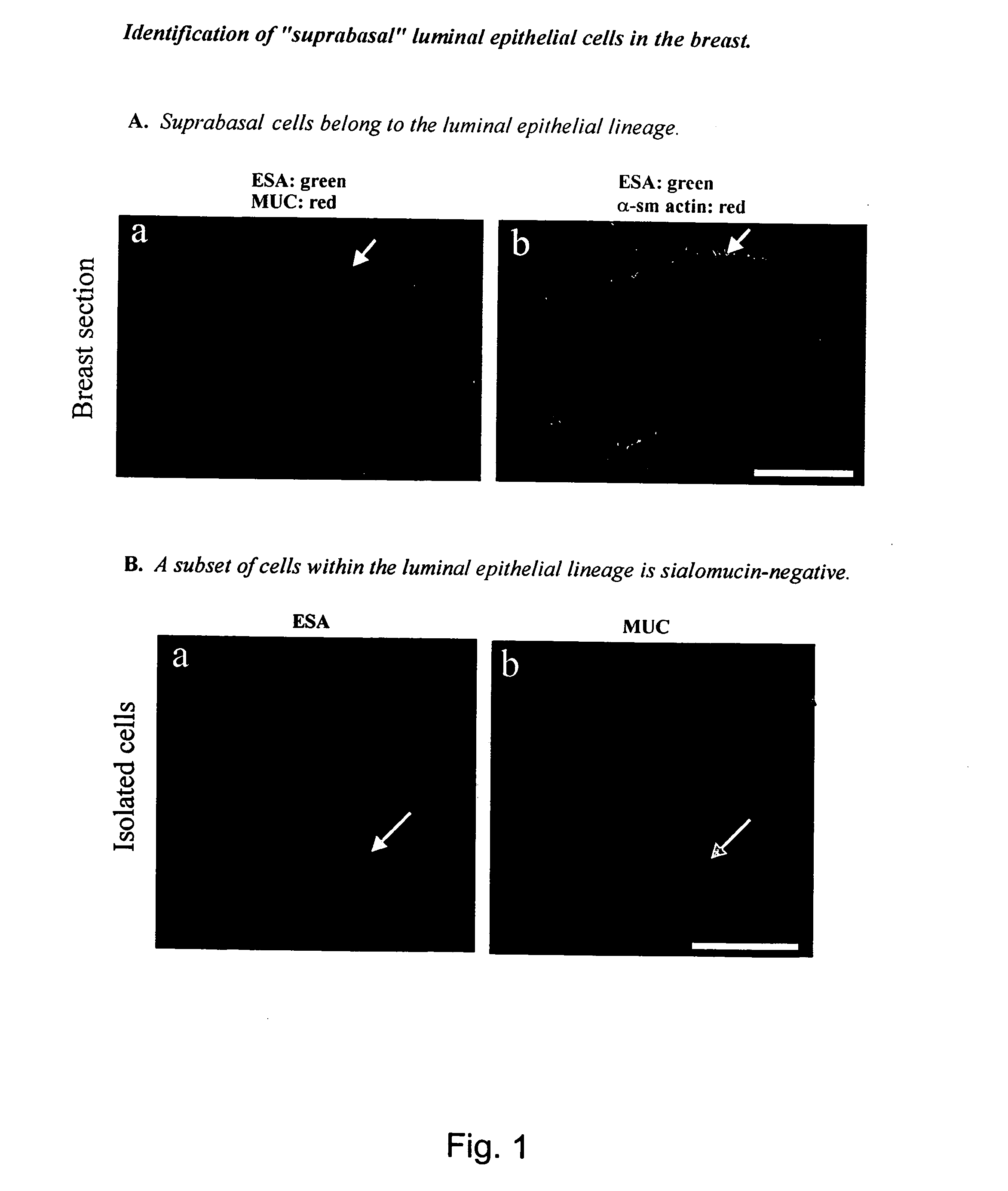
Identification of “Suprabasal” Luminal Epithelial Cells in the Breast

[0105] In culture, a putative “stem” cell of the human breast was defined based on a positive staining for the luminal epithelial marker ESA and a negative or weakly positive staining for sialomucin (MUC) (Stingl et al. 1998). To investigate if such a candidate stem cell could be identified in human breast in vivo, the present inventors double-stained histological sections of normal human breast tissue with epithelial-specific antigen (ESA) and sialomucin (MUC).

[0106] In general, immunocytochemistry and confocal microscopy was performed as follows. Normal human breast tissue was obtained as biopsies from patients undergoing reduction mammoplasty for cosmetic reasons. The use of human material has been reviewed by the Regional Scientific-Ethical Committees for Copenhagen and Frederiksberg, Denmark and approved with reference to (KF) 01-161 / 98. The tissue was frozen in n-hexan (Merck, Darmstadt, Germany) and mount...

example 2

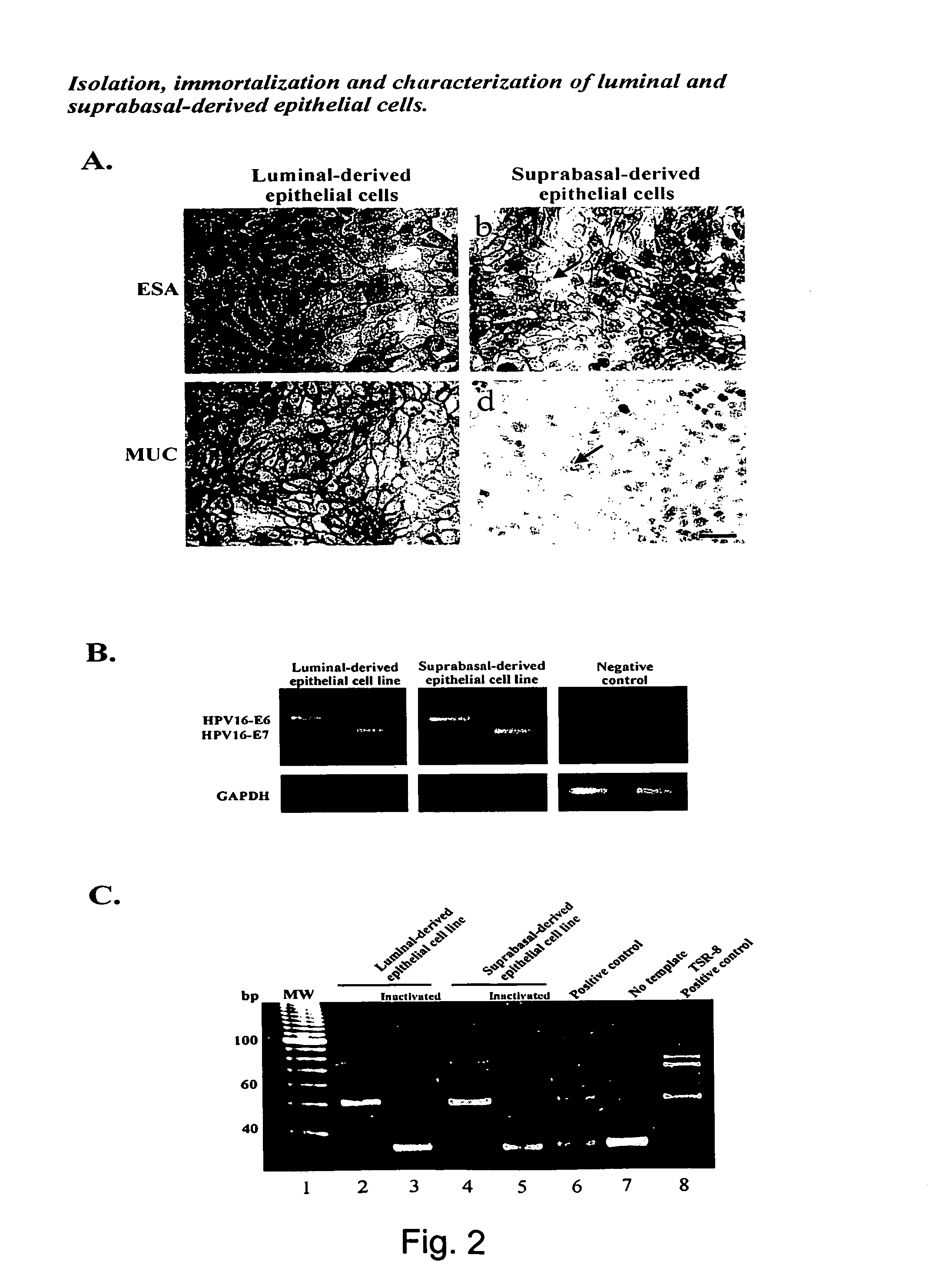
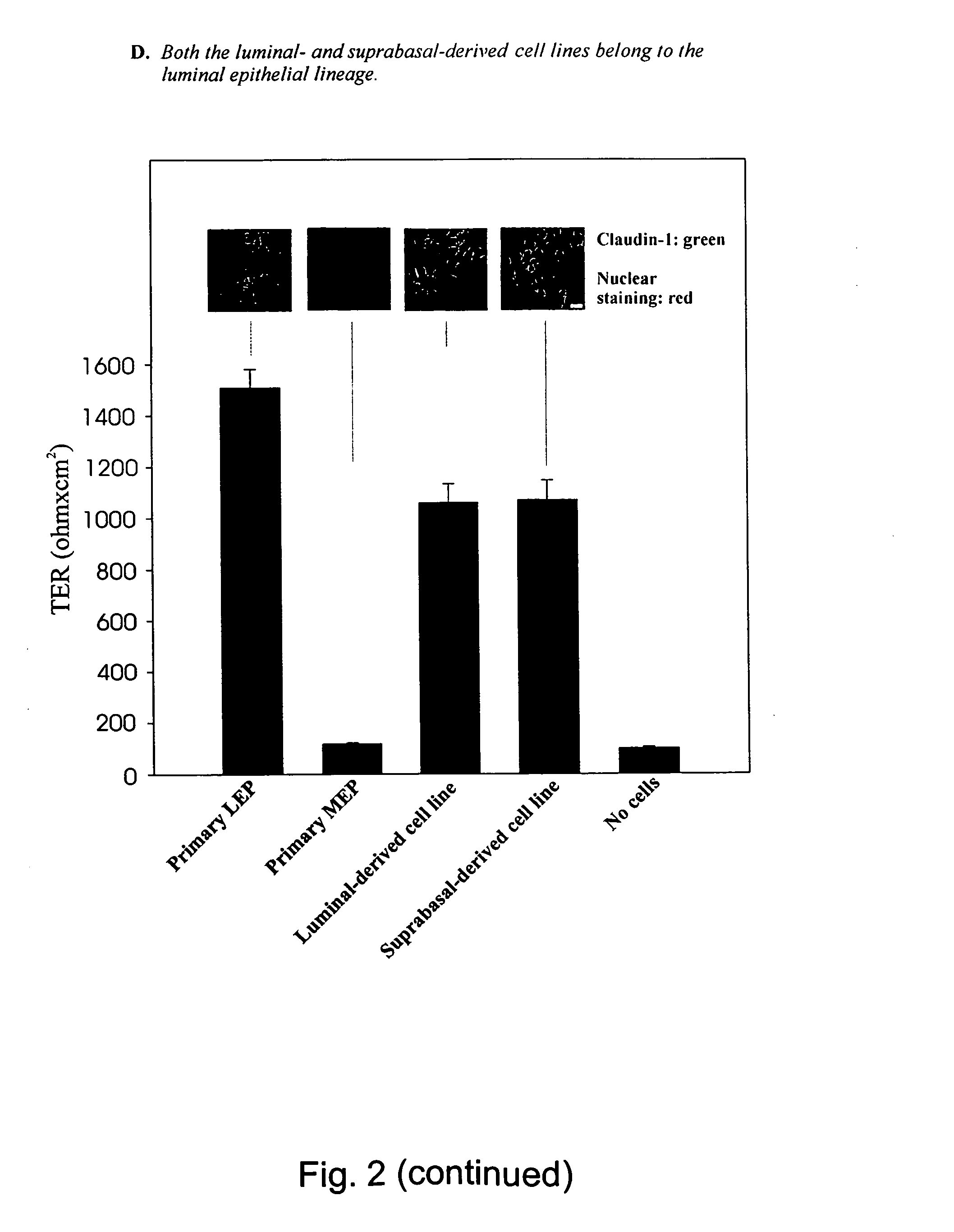
Isolation, Immortalization and Characterization of Luminal and Suprabasal-Derived Epithelial Cells.

[0110] In order to show that the cells described in example 1 indeed have stem cell properties, the cells were isolated by immunomagnetic sorting and characterized.

[0111] Briefly, the luminal epithelial cells were purified from two consecutive sialomucin-columns and the suprabasal epithelial cells were purified as the flow-through from a sialomucin-column which was later retained in an ESA-column. To generate cell lines, the present inventors immortalised both populations with an E6 / E7 construct of HPV16. The resulting established cell lines are referred to below as the luminal and suprabasal-derived epithelial cells, respectively.

Cell Culture

[0112] Breast luminal epithelial cells were generated from primary cultures of biopsies from patients undergoing reduction mammoplasty for cosmetic reasons. The tissue was prepared as previously described (Péchoux et al. 1999). Briefly, it w...

example 3

Clonal Cell Lines of the Suprabasal-Derived Epithelial Cell Line are Multipotent

[0128] Clonal cultures were established and double-stained for keratin K18 (luminal marker) and K14 (myoepithelial marker) as described in example 1 by using antibodies against keratin K18 (F3006; Trichem Aps, Denmark) and keratin K14 (LL002, NovoCastra, Newcastle upon Tyne, UK) as primary antibodies, se also Table 4.

[0129] Whereas the luminal-derived epithelial cell line did not generate any myoepithelial cells and stained for K18 only, the suprabasal-derived cell line readily formed mixed clones of luminal epithelial and myoepithelial cells (FIG. 3A). The present inventors found that these myoepithelial cells represented a primitive level of myoepithelial differentiation because + cells were also precursor cells of a lineage-restricted progeny within the luminal compartment, they could further mature within this compartment to differentiated cells. Sialomucin-expressing cells were eliminated by rete...


PUM

PropertyMeasurementUnit
pore sizeaaaaaaaaaa
volumeaaaaaaaaaa
volumeaaaaaaaaaa

Abstract

A method for isolating cells which have a suprabasal position and expresses epithelial specific antigen but no sialomucin is provided. The isolated cells are shown to share many of the properties expected of a mammary gland stem cell. Three permanent cell lines that are capable of proliferating and capable of differentiating into cells of mammary gland luminal epithelial and myoepithelial cell lineages were established. Such cells form elaborate branching structures resembling uncultured terminal duct lobular units both by morphology and marker expression both in vitro and in vivo. Evidence is provided that these keratin K19 expressing cells most probably is the cells in which breast cancer arises. Thus they constitute a model not only for the developing breast, but also for the development of breast cancer. Also disclosed are the uses of such isolated cells or the cell lines as a model system of the mammary gland for pharmacological studies and their uses in tissue repair or transplantation.

Description

FIELD OF INVENTION [0001] The present invention relates to the isolation of a new at least bi-potent cell type from luminal epithelial cells of a mammary gland and its establishment as an immortalised cell line which is capable of proliferating and capable of differentiating into cells of mammary gland luminal epithelial and myoepithelial cell lineages. The invention furthermore relates to the uses of the isolated cells or the cell lines as a model system of the mammary gland and to the uses in tissue repair or transplantation. GENERAL BACKGROUND [0002] Understanding how the normal human breast develops and which cell compartment becomes neoplastic by necessity is dependent on the isolation of relevant cells as the true targets of human breast carcinogenesis and progression. More than two decades ago it was proposed that human breast cancer originates from the luminal epithelial lineage within the terminal duct lobular units (TDLU), a basic mammary structure consisting of a branchin...

Claims


Application Information

Patent Timeline
Patent Type & Authority Applications(United States)
IPC IPC(8): A61K35/12C12N5/074G01N33/50
CPCA61K35/12C12N5/0631C12N2503/02G01N2333/4742C12N2510/04G01N33/5011G01N33/5017C12N2510/00
Inventor PETERSEN, LONEPETERSEN, OLEGODJONSSON, THORARINNVILLADSEN, RENEBISSELL, MONA
Owner UNIVERSITY OF COPENHAGEN
Why Eureka
Try Eureka