
Image-based method for detection and removal of small fragments in segmented three-dimensional volumes

Active Publication Date: 2005-11-24
SIEMENS MEDICAL SOLUTIONS USA INC
View PDF3 Cites 13 Cited by
  • Summary
  • Abstract
  • Description
  • Claims
  • Application Information

AI Technical Summary

Benefits of technology

[0007] The present invention is directed to a method for detecting and removing small isolated fragments in a 3D segmented volume. The 3D segmented volume is projected onto several 2D images from different viewing directions. Isolated 2D fragments are detected in the 2D images. Corresponding 3D frag

Problems solved by technology

The side effect of generating small fragments representing noisy structures surrounding larger target objects within the desired range of interest is undesirable, but often unavoidable, when segmenting three-dimensional tomographic data.
A major drawback of this brute force approach is that both regions belonging to the large and small fragments are required to be traversed and their size computed.
Therefore, it is very processing intensive, time consuming and impractical for large amounts of data.
Manual picking and removal of such fragments is only feasible if the number of fragments is not many, otherwise, this becomes a tedious and time consuming approach.
Unfortunately, large tomographic medical data tend to have a large number of such undesired fragments and require longer processing time.
It is therefore neither practical to remove noise fragments by brute force automatic methods, nor by picking each fragment manually.

Method used


Image

  • Image-based method for detection and removal of small fragments in segmented three-dimensional volumes
  • Image-based method for detection and removal of small fragments in segmented three-dimensional volumes
  • Image-based method for detection and removal of small fragments in segmented three-dimensional volumes

Examples


Embodiment Construction

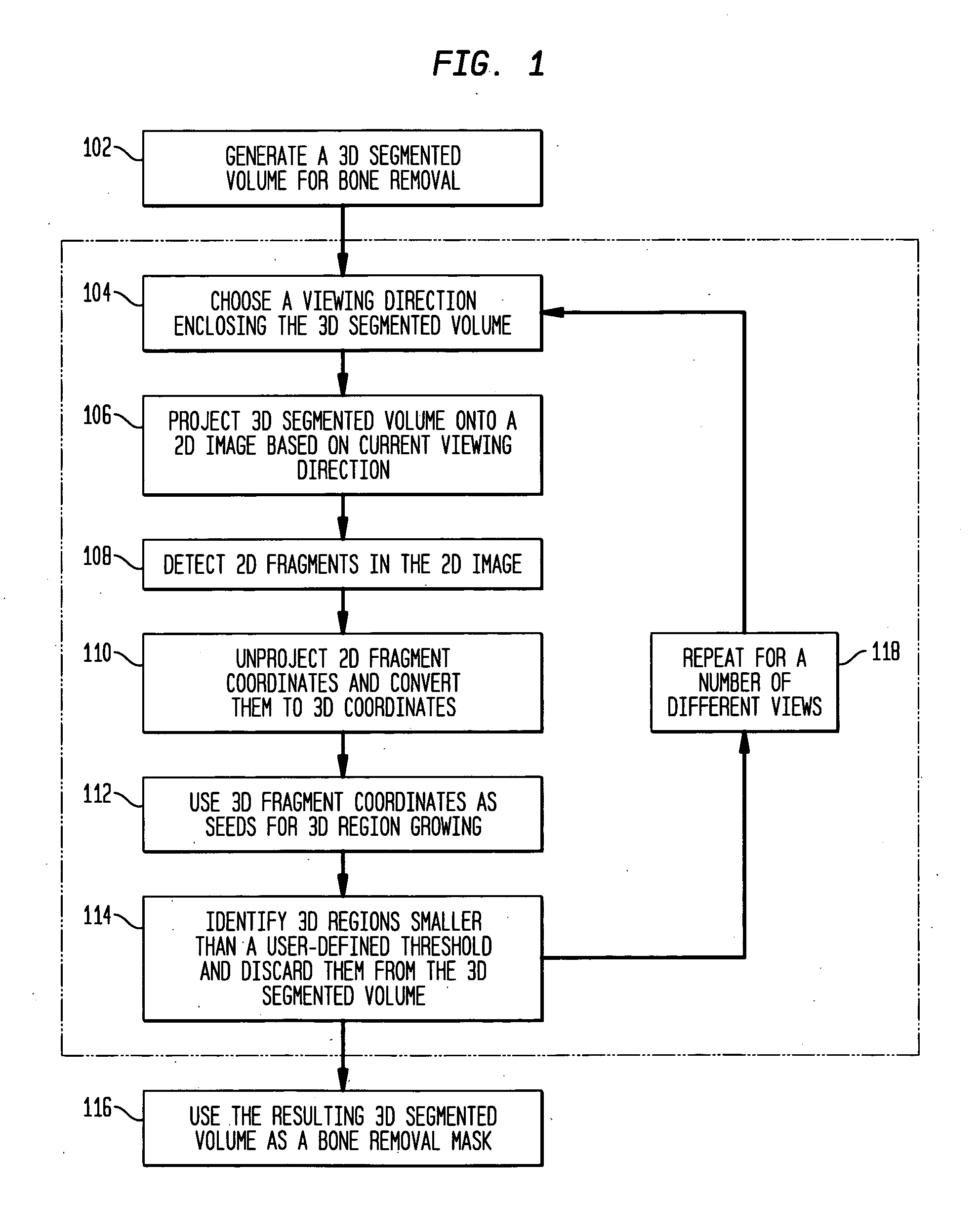
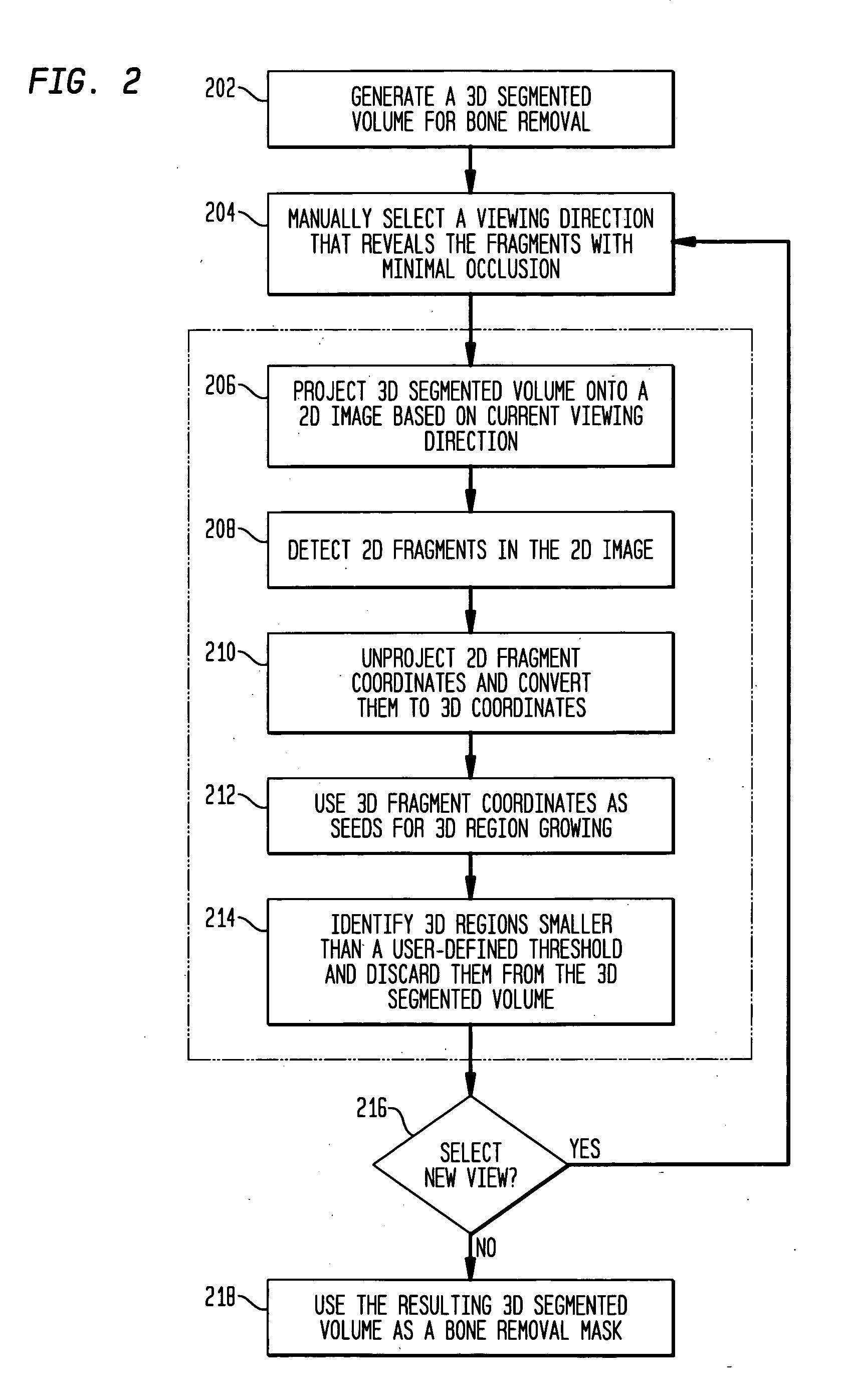
[0012] The present invention is directed to a method for the removal of small isolated fragments in segmented three-dimensional volumes of tomographic medical data. In accordance with the present invention, the time-consuming manual picking of 2D point coordinates used for ray picking and 3D region growing is replaced by an image-based approach that detects 2D point coordinates corresponding to fragments in a 2D volume image that is rendered automatically. To improve the efficacy of this image-based method, several 2D volume rendered images from different viewing directions are generated and seed points are identified in each one of them. The efficacy of fragment detection is further improved by partitioning the volume into sub-sections, rendering each 3D subsection to several 2D images from different viewing directions, and detecting 2D seed point coordinates for each sub-section. Alternatively, the viewing directions can be selected by the user manually. One application for the me...


PUM

PropertyMeasurementUnit
Volumeaaaaaaaaaa

Abstract

A method for detecting and removing small isolated fragments in a 3D segmented volume is disclosed. The 3D segmented volume is projected onto several 2D images from different viewing directions. Isolated 2D fragments are detected in the 2D images. Corresponding 3D fragments are found in the 3D volume by unprojecting corresponding detected 2D fragment locations. The unprojected detected 2D fragment locations are used as seed points for region growing of isolated 3D fragments. Any of the 3D fragments having a volume size below a user-defined threshold are discarded.

Description

CROSS REFERENCE TO RELATED APPLICATIONS [0001] This application claims the benefit of U.S. Provisional Patent Application Ser. No. 60 / 573,961, filed May 24, 2004, which is incorporated by reference in its entirety.FIELD OF THE INVENTION [0002] The present invention relates to image-based segmentation, and more particularly to a method for automatically detecting and removing small isolated fragments in segmented three-dimensional volumes of large tomographic medical data. BACKGROUND OF THE INVENTION [0003] The side effect of generating small fragments representing noisy structures surrounding larger target objects within the desired range of interest is undesirable, but often unavoidable, when segmenting three-dimensional tomographic data. For example, when using a global threshold method to segment a target object in a grayscale volume, each voxel is classified into object and background determined by whether a voxel belongs to a selected group of intensity threshold values. When n...

Claims


Application Information

Patent Timeline
IPC IPC(8): C12Q1/68G01N33/48G01N33/50G06F19/00G06T5/00G06T15/00G06T15/40
CPCG06K9/346G06K2209/05G06T7/0081G06T2200/04G06T2207/10081G06T2207/30008G06T5/005G06T15/08G06T7/11G06V10/273G06V2201/03G06T5/77
Inventor PALADINI, GIANLUCAYU, DAPHNE
Owner SIEMENS MEDICAL SOLUTIONS USA INC
Features
  • Generate Ideas
  • Intellectual Property
  • Life Sciences
  • Materials
  • Tech Scout
  • Unparalleled Data Quality
  • Higher Quality Content
  • 60% Fewer Hallucinations
Learn More