

Detection of measurement of antibodies to antigenic proteins in biological tissues or samples
a technology of antigenic protein and detection method, which is applied in the field of biotechnology and medical diagnostics, can solve problems such as the difficulty of distinguishing the labeled induced antibody-antigenic protein complex
- Summary
- Abstract
- Description
- Claims
- Application Information
AI Technical Summary
Problems solved by technology
Method used
Image

Examples
example 1
Detection of Experimentally Formed Antigen and Monoclonal Antibody Immune Complex by Intercolation of Labeled Antigen into the Immune Complex
[0027] The immune complex of antigenic protein (infliximab, 15.3 ug / mL) and induced murine monoclonal antibody to the antigenic protein (5.1 ug / mL, 3:1 molar ratio) was experimentally formed in normal human serum. At these specified concentrations, the induced monoclonal antibody was completely bound by the excess antigenic protein and not detectable using current in vitro assay formats.
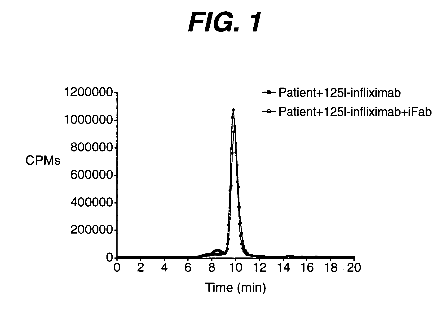
[0028] The CPM chromatogram in FIG. 1 shows that the retention time of 125I-labeled antigenic protein (infliximab) is approximately 16.4 minutes, which is characteristic of the protein's size and shape. The retention time remains relatively constant when the HPLC column, flow parameters and mobile phase buffer, are left unchanged.
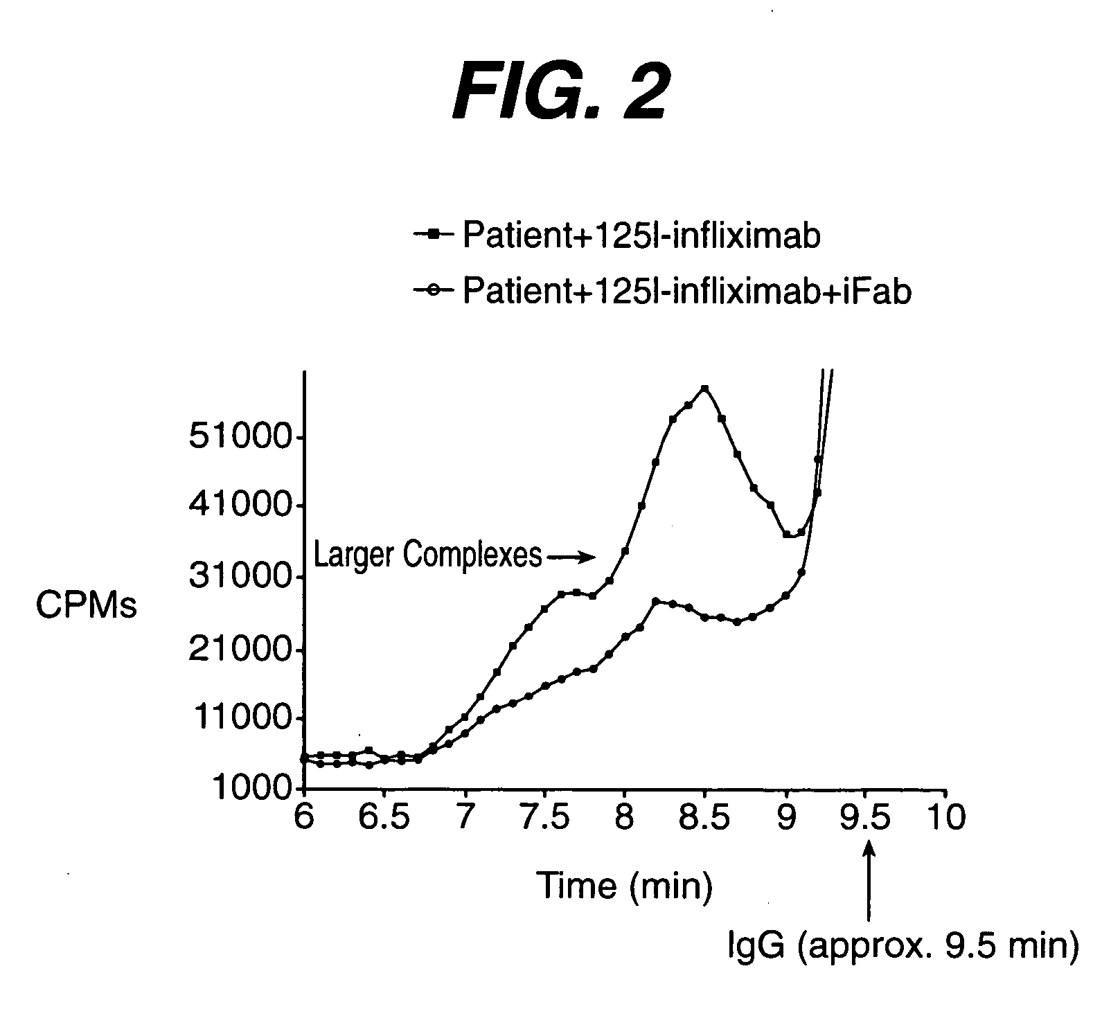
[0029] The immune complex of an antigenic protein and its induced antibody is larger in size than each of the individual component....
example 2
Detection of Experimentally Formed Antigen and Polyclonal Antibody Immune Complex by Intercolation of Labeled Antigen into the Immune Complex
[0032] The immune complex of antigenic protein (infliximab, 15.3 ug / mL) and induced monkey polyclonal antibody to the antigenic protein (5.1 ug / mL, 3:1 molar ratio) was experimentally formed in normal human serum. At these specified concentrations, the induced polyclonal antibody was completely bound by the excess antigenic protein and not detectable using current in vitro assay formats.
[0033] To demonstrate that induced polyclonal antibody can be detected through radiolabeled antigenic protein, serum containing immune complex of infliximab and induced polyclonal antibody against infliximab was incubated in the presence of excess 125I-labeled infliximab for 1 hour at 37 degrees. FIG. 5 shows the CPM chromatogram with peaks at 24.8, 16.8, 14.4 and 13.2 minutes. These are retention times characteristic of free 125I not associated with protein (...
example 3
Detection of Infliximab and Induced Anti-Infliximab Antibody Immune Complexes in Patient Serum
[0034] Serum samples were taken from patient A at week 0 and week 28 after the initiation of infliximab treatment (8 weeks after the latest infliximab infusion). Both were determined by double antigen EIA analysis to be negative for induced antibodies to infliximab. No circulating infliximab was detectable using a validated ELISA in either sample.
[0035] The serum was incubated with approximately 15 μg / mL of 125I-labeled infliximab for at least one hour at 37 degrees on a shaking platform. For serum sample taken at week 0 (FIG. 6A), a single peak was detected at 16.4 minutes, the retention time of uncomplexed 125I-labeled infliximab. There is no significantly visible peak at less than 16.4 minutes (the percentage of the area under the chromatogram of retention time less than 16.4 minutes over the total chromatogram area is approximately 11.6%), which suggests that no complex with higher mo...
PUM
| Property | Measurement | Unit |
|---|---|---|
| size exclusion column | aaaaa | aaaaa |
| size exclusion column | aaaaa | aaaaa |
| molecular weight | aaaaa | aaaaa |
Abstract
Description
Claims
Application Information