Method and apparatus for screening for retinopathy

a technology of retinopathy and screening method, applied in the field of screening method and screening apparatus for retinopathy, can solve the problems of diabetes, vision loss, major health threat to diabetics, and failure to recognize and treat diabetic retinopathy

Active Publication Date: 2006-04-13
UNIVERSITY OF PITTSBURGH
View PDF3 Cites 29 Cited by
  • Summary
  • Abstract
  • Description
  • Claims
  • Application Information

AI Technical Summary

Benefits of technology

[0027] It is a further object of this invention to provide a method and apparatus for screening for retinopathy that can yield an immediate clinical assessment.

Problems solved by technology

Diabetic retinopathy, which is a pathological disorder of the retina caused by diabetes, is a major health threat to diabetics.
The most common cause of vision loss in diabetics is the failure to recognize and treat diabetic retinopathy.
When uncontrolled, angiogenesis can cause damage to normal retinal tissues because the new vessels are fragile and hemorrhage easily.
In addition, ischemic changes promote increased permeability of the retinal blood vessels, which leads to a swelling and thickening of the central portion of the retina, a condition called macular edema (the macula is an area near the center of the retina), and reduced visual function.
Unlike macular edema, there is currently no known quantitative screening method for retinal ischemia.
This is due, in part, to the fact that the midperipheral portion of the retina is poorly visualized by routine clinical exams and most retinal photography systems.
As a result, screening programs based on clinical examinations have a high no show rate, and many cases of diabetic retinopathy are not diagnosed until proliferative retinal changes are extensive.
Such systems have several disadvantages, including delays in determining patient status, since images must be transmitted to and reviewed by a remote clinician (such delays create a potential for loss of follow-up with the patient), the high manpower costs required to implement the systems, the limitations imposed by media opacities that affect the quality of the transmitted images, and the fact that such systems still rely on subjective clinical examinations and, as a result, present the same problems described above.
A fluorescein angiogram, however, is an invasive procedure that requires dilation of the eye, the injection of dye into the patient and highly specialized photography.
As a result, fluorescein angiograms present the potential for numerous complications.
These techniques have several disadvantages associated with them.
In particular, visual field testing is subjective, time consuming, and relies on the active and accurate participation by the patient, and multi-focal electrophysiology is costly, time consuming, and requires dilation and significant patient cooperation.

Method used


Image

  • Method and apparatus for screening for retinopathy
  • Method and apparatus for screening for retinopathy
  • Method and apparatus for screening for retinopathy

Examples


Embodiment Construction

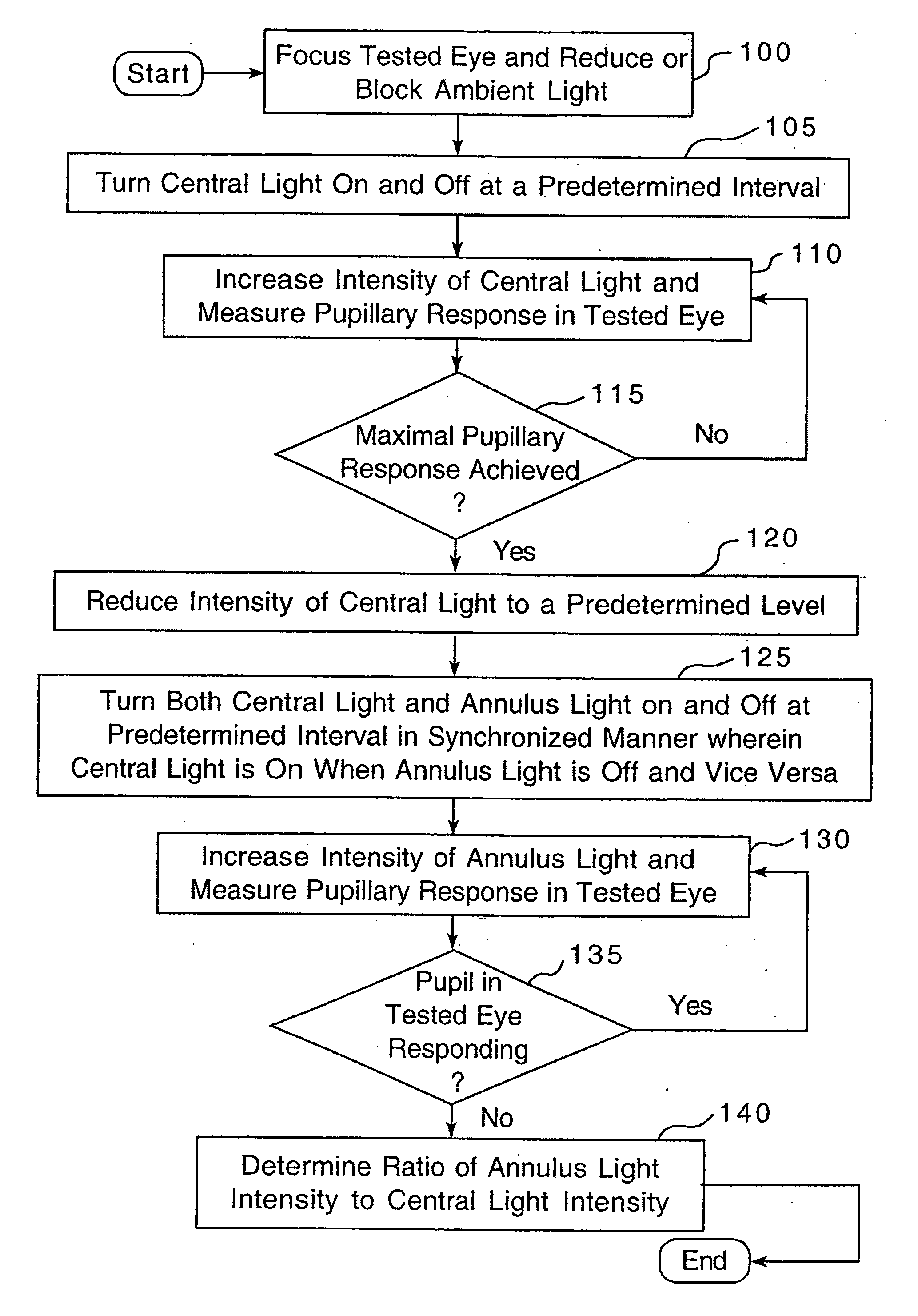
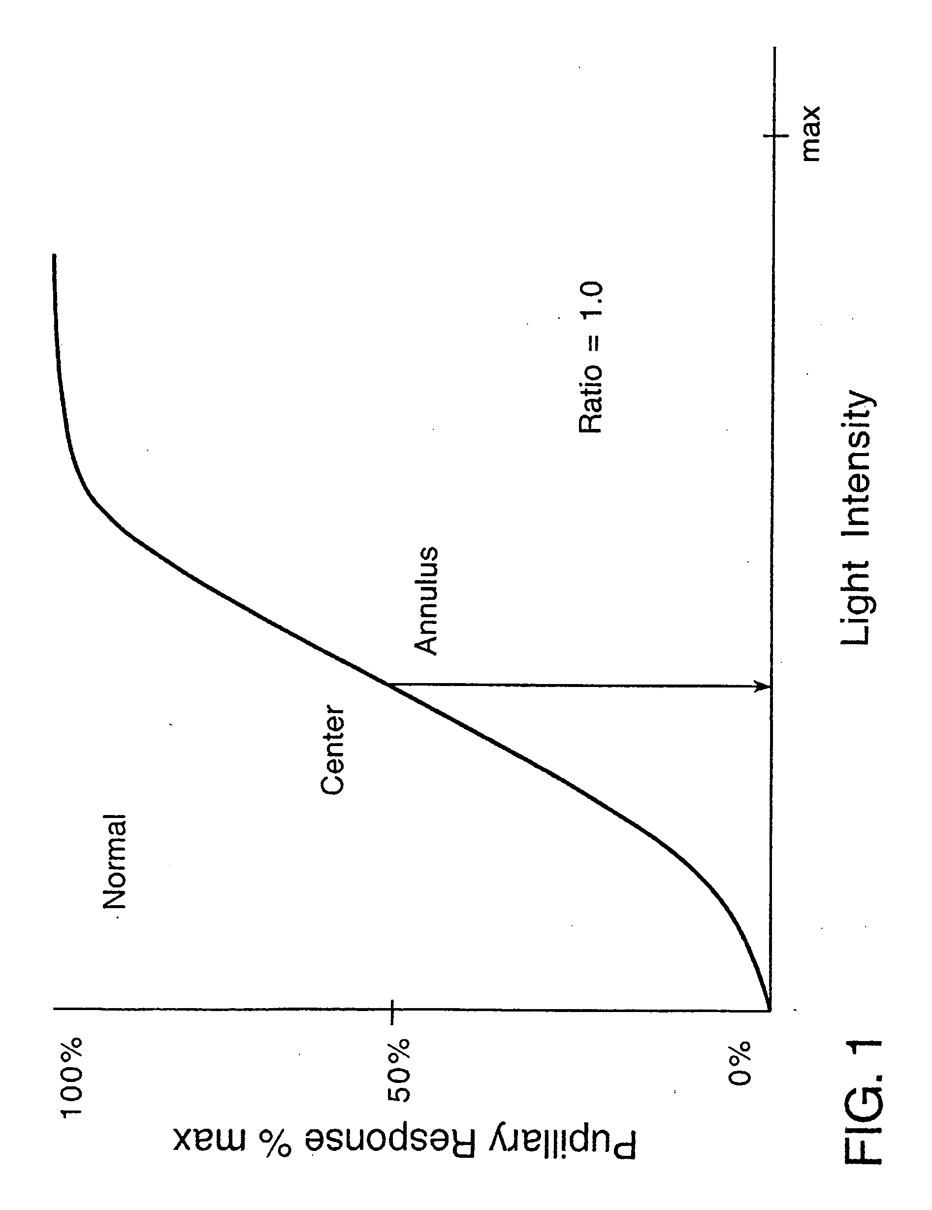
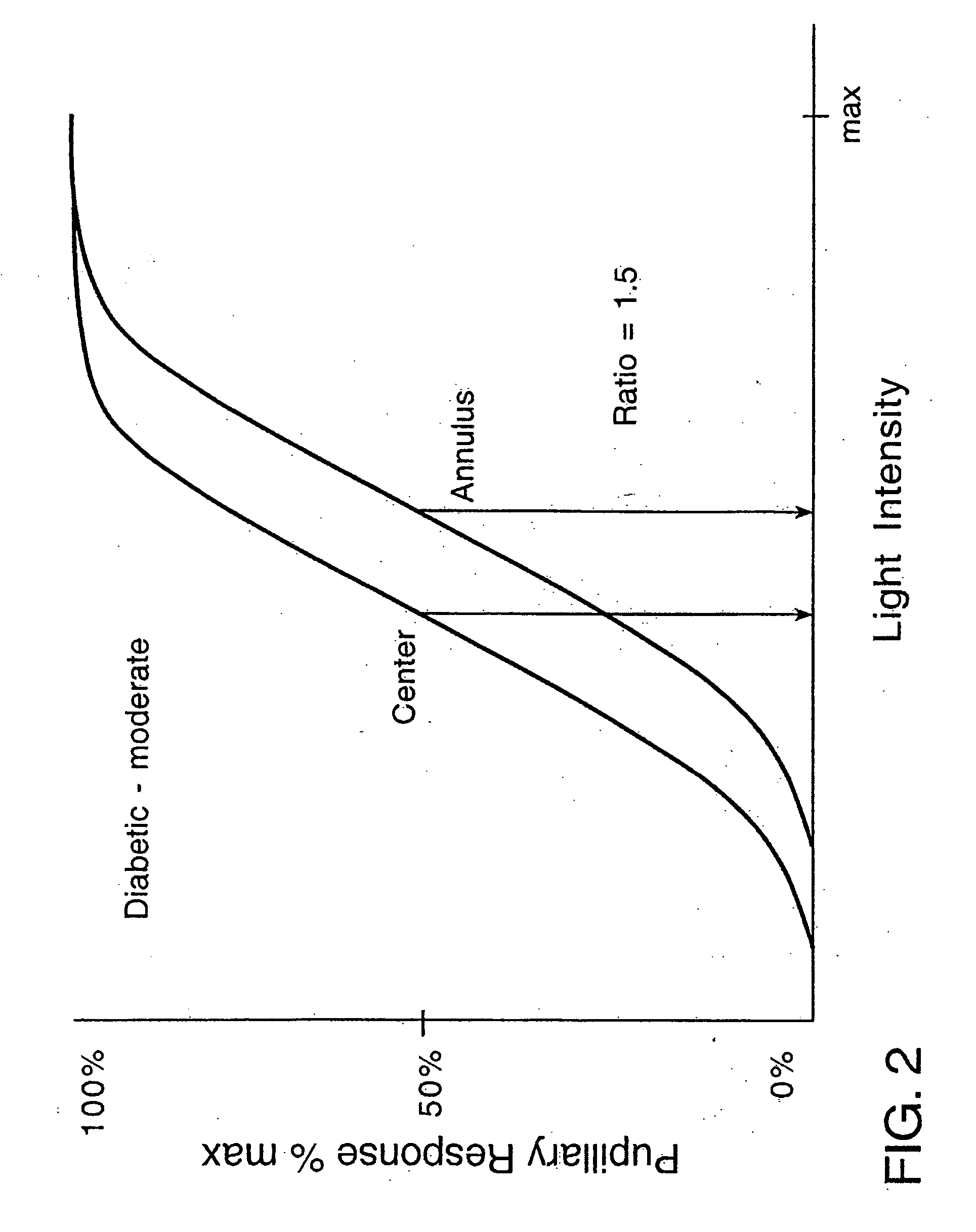
[0038] In healthy people having normal vision, different portions or areas of the retina, such as the central portion and the midperipheral portion, are generally equally sensitive to light. As a result, light of a particular intensity that is independently incident upon each of those different regions will produce generally the same pupillary response, i.e., the pupil will constrict to the same degree. In contrast, as discussed elsewhere herein, certain eye diseases cause reduced sensitivity to light in particular retinal areas as compared to others. For example, in diabetic retinopathy, the sensitivity of both the central and midperipheral portions of the retina to light are reduced, with the sensitivity of the midperipheral portion being reduced to a much greater degree.

[0039] As is known, the sensitivity of the retina to light may be observed by measuring the pupillary response to light. FIG. 1 is a graph of pupillary response, as a percentage of maximum pupil constriction, ver...


PUM


Abstract

A method of screening for retinal disease including directing a first light at a first portion of the retina, directing a second light at a second portion of the retina, measuring a first pupillary response of the eye as a result of the first light and a second pupillary response as a result of the second light, and generating an indication of a severity level of the retinal disease using the first pupillary response and the second pupillary response. Also, an apparatus that implements this method including a light source for directing a first light at a first retinal portion and a second light at a second retinal portion, a pupil measuring device for measuring the pupillary response of the eye as a result of the first light and second light, a processor in electronic communication with the pupil measuring device, and a memory in electronic communication with the processor.

Description

CROSS-REFERENCE TO RELATED APPLICATION [0001] The instant application claims priority from U.S. Provisional Patent Application Ser. No. 60 / 616,160 filed Oct. 5, 2004, the disclosures of which are incorporated herein by reference.BACKGROUND OF THE INVENTION [0002] 1. Field of the Invention [0003] The present invention relates to a method and apparatus for screening for retinopathy, such as diabetic retinopathy, and in particular to a method and apparatus for providing an objective indication of the severity of retinal disease and disease features, such as diabetic retinopathy and retinal ischemia, using pupillometry and a plurality of light stimuli. [0004] 2. Description of Related Art [0005] There is an epidemic of diabetes in this country. Diabetic retinopathy, which is a pathological disorder of the retina caused by diabetes, is a major health threat to diabetics. It is estimated that more than 80% of diabetics will develop diabetic retinopathy to some extent, with a large percent...

Claims


Application Information

Patent Timeline
Patent Type & Authority Applications(United States)
IPC IPC(8): A61B3/10A61B3/00
CPCA61B3/0008A61B3/12A61B3/112A61B3/10
Inventor GORIN, MICHAEL B.
Owner UNIVERSITY OF PITTSBURGH
Why Eureka
Try Eureka