Treatment of age-related macular degeneration

a technology treatment method, which is applied in the field of age-related macular degeneration treatment, can solve the problems of central vision distortion, obscuring the ability of a physician, and severely affecting the patient's vision at the highest resolution, and achieves reliably accurate directional emission and placement. accurate

Inactive Publication Date: 2006-04-13
XOFT INC +1
View PDF5 Cites 126 Cited by
  • Summary
  • Abstract
  • Description
  • Claims
  • Application Information

AI Technical Summary

Benefits of technology

[0014] An important advantage of delivering ionizing radiation from the back of the eye is that a therapeutic dose can be given to the lesion without damaging structures of the eye, i.e. the lens, retina and optic nerve. The membranes at the back of the eye are relatively radioresistant.
[0016] Infrared radiation can be used advantageously in location of the probe. IR can be directed in from the front of the eye, invisible to the patient and thus not uncomfortable to the patient, and this radiation will penetrate through the retina, choroid and sclera to the sensors on the device.
[0019] Filtration of the x-ray beam from the miniature tube can be put in place if needed. This can greatly reduce low-energy emissions (i.e. “hardening” the beam), thus reducing dose in the near field, i.e. the sclera, where radiation is not desired. This in combination with standoff of the source after placement and adjustment of voltage, enables optimizing dose to the choroid, sclera and retina.
[0020] It is important that the probe be immobilized during treatment. Several methods can be used. The “bump” on the probe device described above can be inflatable, thus being inserted flattened but inflated (with liquid) when near the macula. Further inflation can help hold the probe against the adjacent tissues (and also stands the source off from the tissue as described below). Another inflatable balloon can be at the back side of the probe, and a further balloon(s) can be located on the probe proximal to the x-ray source for further immobilization of the probe.

Problems solved by technology

The term “wet” refers to the fact that these abnormal blood vessels leak and damage the macula, causing central vision distortion.
Thus, the highest resolution vision of the patient is severely compromised or lost.
Approximately ten percent of AMD cases comprise wet AMD, and this is the type which can lead to blindness.
In the case of classic AMD, the new blood vessels remain essentially intact, while in occult AMD the blood vessels leak, form a somewhat amorphous mass, and obscure the ability of a physician to see the vessel through an ophthalmoscope.
Thus, any treatment aimed specifically at these vessels, requiring visual identification and location of the vessels, fails for occult AMD.
Results of both TTT and PDT have been less than ideal.
In addition, the cost for this treatment has been extremely high and its cost effectiveness is in question.
Thus, patients will probably need three to four treatments per year for this therapy to be effective, and the treatment basically preserves vision rather than improves visual acuity.
External beam treatment is difficult and generally limited to highly specialized practitioners, with capital cost for equipment very high.
This treatment is difficult for several reasons, including inability to produce a very small beam specific to the target, location of the beam precisely on the target, radiation damage to other structures, including brain tissue, and avoidance of irradiating the optic nerve and retna.
In addition to other limitations discussed above, the use of isotopes is not ideal.
They are extremely limited as to choices of specific activity of the isotopes, as well as energy.

Method used


Image

  • Treatment of age-related macular degeneration
  • Treatment of age-related macular degeneration
  • Treatment of age-related macular degeneration

Examples


Embodiment Construction

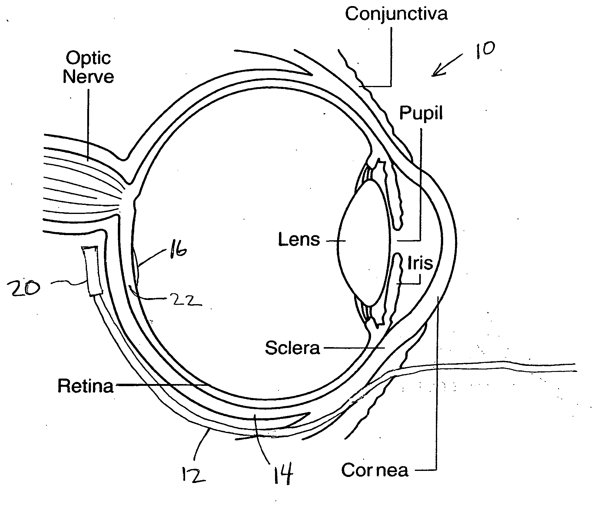
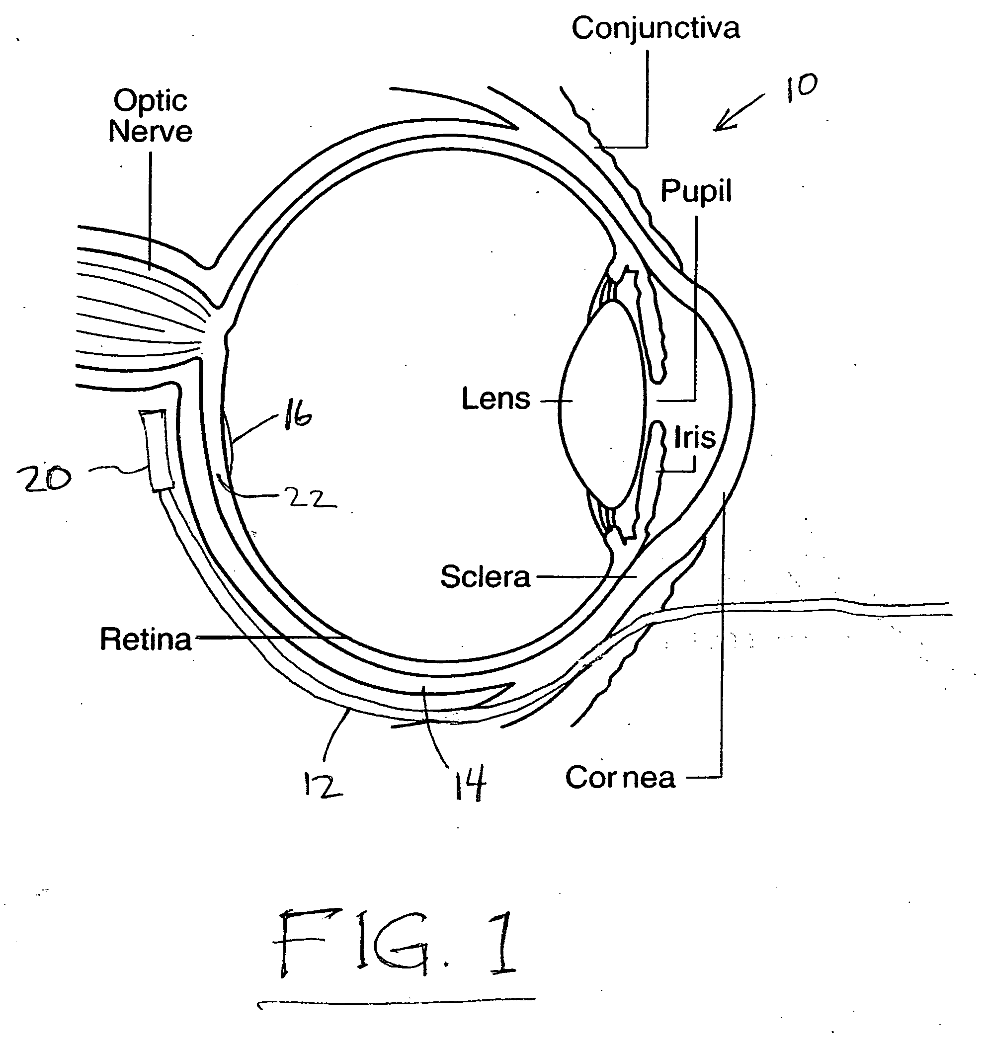
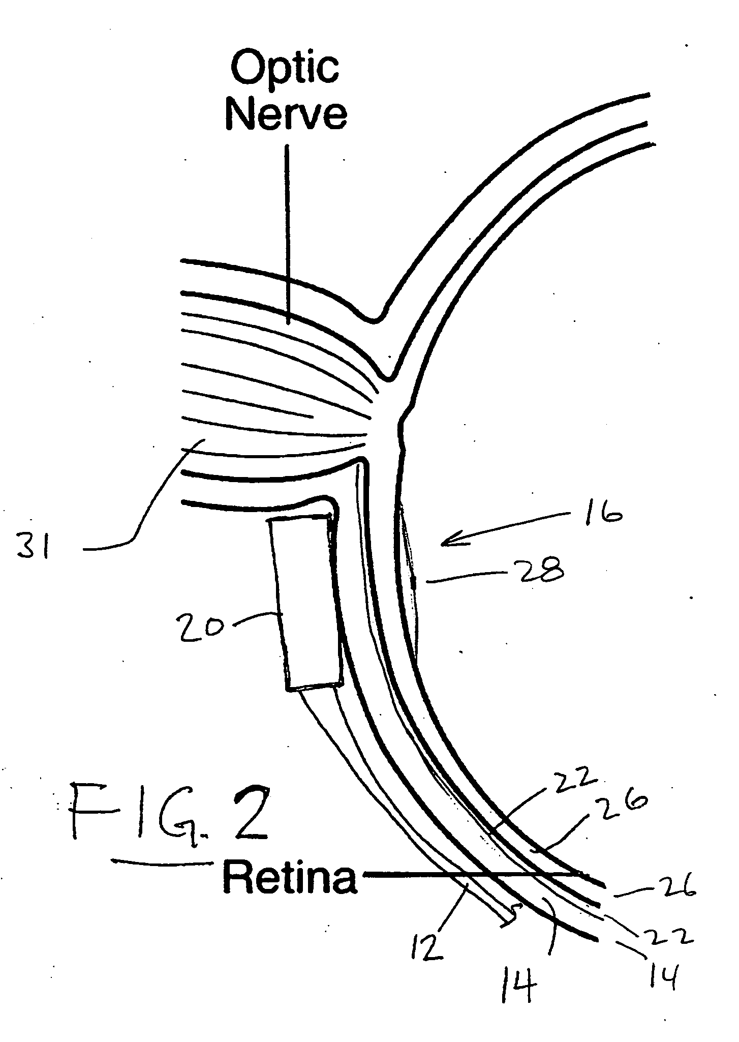
[0047]FIG. 1 of the drawings shows a patient's eye 10, generally in plan view and cross section, and schematically indicates a probe or catheter device 12 inserted around the globe of the eye, along the surface of the sclera 14. The macula, or macular region, a region of the retina of the eye, is shown at 16. The probe or catheter device 12, which is shown schematically, includes a switchable x-ray source 20 at its distal end. An applicator or guide device may be included, to be inserted around the eye and through which the catheter is inserted. The source 20 preferably comprises an x-ray tube which emits radiation when switched on and which optionally can be varied as to current and voltage, emitting a side-looking directional radiation toward the macula 16, through the sclera 14 and the choroidal membrane or layer immediately behind the macula. The source could be an isotope, with controlled shielding if desired. Accurate positioning of the x-ray source 20 is an important aspect o...


PUM


Abstract

Age-related macular degeneration is treated by radiation delivered from a miniature x-ray tube inserted via a catheter around the globe of the eye, to a position behind the macula. The process can employ an applicator with several parallel guides, inserted around the eye to receive the catheter with x-ray tube (or an isotope) at a matrix of different positions. Methods are described for properly locating the catheter and x-ray tube, using illumination on the catheter and viewing through the front of the eye, or sensors on the catheter and a scanned beam shone from the front of the eye. Fluorescent material excited by x-rays can also be used. Also described are methods and devices for immobilizing the probe once properly located in the eye, for standoff of the x-ray tube from the target tissue, and for achieving prescription radiation dose in the choroid while eliminating dose to adjacent tissues. The x-ray treatment can be enhanced using a radiosensitizing drug, and can be combined with PDT.

Description

[0001] This application is a continuation-in-part of application Ser. No. 10 / 635,421, filed Aug. 6, 2003.BACKGROUND OF THE INVENTION [0002] This invention concerns treatment of wet age-related macular degeneration (AMD) in the eye, and in particular concerns use of radiation to treat such macular degeneration, the radiation being administered from behind the sclera. Treatment of ocular tumors also forms a part of the invention. [0003] Wet age-related macular degeneration has been the leading cause of blindness in the United States, and a leading cause in the world. It is characterized by the growth of abnormal blood vessels from the choroidal membrane at the back of the eye, in the macular area of the retina including the fovea and immediately surrounding regions. This is called “choroidal neovascularization” (CNV). The term “wet” refers to the fact that these abnormal blood vessels leak and damage the macula, causing central vision distortion. Thus, the highest resolution vision of...

Claims


Application Information

Patent Timeline
Patent Type & Authority Applications(United States)
IPC IPC(8): A61N5/10A61B6/00A61B19/00A61KA61N5/00A61N5/06
CPCA61B2019/5206A61F9/0079A61N5/0601A61N5/062A61N5/1001A61N5/1017A61N2005/0651A61N2005/0659A61N2005/1022A61N2005/1098A61B2090/306
Inventor FORMAN, MICHAELLOVOI, PAUL A.SMITH, PETER C.
Owner XOFT INC
Why Eureka
Try Eureka