Systems and methods for viewing medical 3D imaging volumes

Active Publication Date: 2006-05-04
MERATIVE US LP
View PDF99 Cites 76 Cited by
  • Summary
  • Abstract
  • Description
  • Claims
  • Application Information

AI Technical Summary

Benefits of technology

[0013] Yet another embodiment comprises a method of viewing a 3D imaging volume on a display device coupled to a computing system. The display device is configured to concurrently display one or more navigation images and one of a plurality of reformatted images that comprise a plane of the imaging volume along any axis chosen by the view. The method comprises receiving an imaging volume at the computing system, wherein the imaging volume comprises a plurality of voxels arranged in a three dimensional array. The method also comprises displaying on the display device one or more na

Problems solved by technology

While a radiologist or physician interpreting such an imaging volume might choose to view the acquired images, the plane in which the images were acquired may not be optimal for accurate and efficient analysis and interpretation of the scan.
Currently available systems for viewing imaging volumes do not provide a mechanism for tracking which portions of the imaging volume, such as which voxels of the imaging volume, have been displayed on a display device.
As those of skill in the art will recognize, requiring the viewer to determine when all relevant portions of an imaging volume have been viewed introduces the possibility that portions of the imaging volume are not viewed and, thus, features and / or abnormalities expressed in the unviewed portions may not be detected.

Method used


Image

  • Systems and methods for viewing medical 3D imaging volumes
  • Systems and methods for viewing medical 3D imaging volumes
  • Systems and methods for viewing medical 3D imaging volumes

Examples


Embodiment Construction

[0022] Embodiments of the invention will now be described with reference to the accompanying figures, wherein like numerals refer to like elements throughout. The terminology used in the description presented herein is not intended to be interpreted in any limited or restrictive manner, simply because it is being utilized in conjunction with a detailed description of certain specific embodiments of the invention. Furthermore, embodiments of the invention may include several novel features, no single one of which is solely responsible for its desirable attributes or which is essential to practicing the inventions herein described.

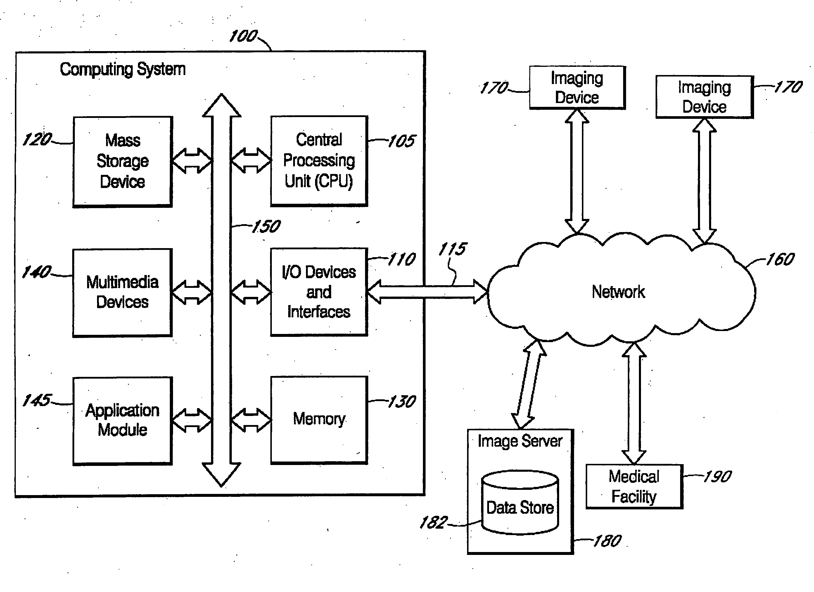
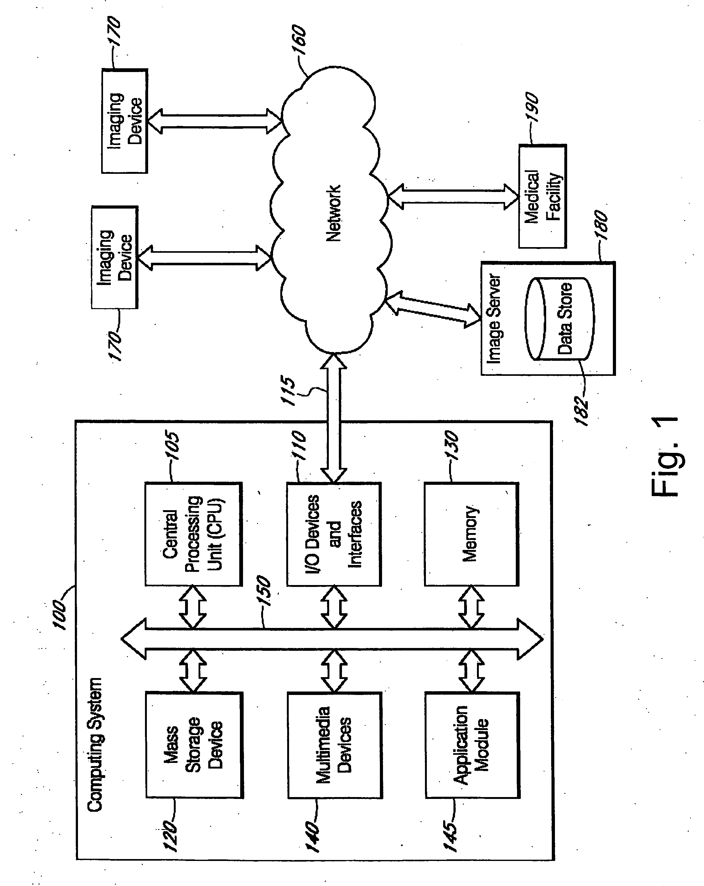
[0023]FIG. 1 is a block diagram of an exemplary computing system 100 in communication with a network 160 and various network devices. The computing system 100 may be used to implement certain systems and methods described herein. The functionality provided for in the components and modules of computing system 100 may be combined into fewer components and mo...


PUM


Abstract

A method of automatically tracking the portions of a 3D medical imaging volume, such as the voxels, that have already been displayed according to use-defined display parameters, notating those portions, and providing the user with information indicating what portions of the imaging volume have been displayed at full resolution.

Description

CROSS-REFERENCE TO RELATED APPLICATIONS [0001] This application claims priority under 35 U.S.C. § 119(e) to U.S. Provisional Application Ser. No. 60 / 625,690, filed on Nov. 4, 2004, which is hereby expressly incorporated by reference in its entirety.BACKGROUND OF THE INVENTION [0002] 1. Field of the Invention [0003] This invention relates to management and viewing of medical images and, more particularly, to systems and methods of tracking which portions of three dimensional imaging volumes have been displayed using predetermined display parameters. [0004] 2. Description of the Related Art [0005] Medical imaging is increasingly moving into the digital realm. This includes imaging techniques that were traditionally analog, such as mammography, x-ray imaging, angiography, endoscopy, and pathology, where information can now be acquired directly using digital sensors, or by digitizing information that was acquired in analog form. In addition, many imaging modalities are inherently digita...

Claims


Application Information

Patent Timeline
IPC IPC(8): G06K9/00
CPCA61B6/5223G06T3/40G06T15/08G06T19/00G06T2210/41G06T2219/008G06T19/20A61B2090/367A61B90/37
Inventor FRAM, EVAN K.REICHER, MURRAY A.
Owner MERATIVE US LP
Why Eureka
Try Eureka