Method to treat collagenous connective tissue for implant remodeled by host cells into living tissue

a technology of host cells and connective tissue, which is applied in the direction of peptide/protein ingredients, prosthesis, ligaments, etc., can solve the problems of minor foreign body reaction, infection, pyrogenic shock or major foreign body reaction of the recipient, and denatured and slightly toxic to the recipien

Inactive Publication Date: 2006-06-15
CHEUNG DAVID T
View PDF2 Cites 5 Cited by
  • Summary
  • Abstract
  • Description
  • Claims
  • Application Information

AI Technical Summary

Benefits of technology

[0019] There is, therefore, provided according to the present invention, a method for processing collagenous connective tissue from an animal donor source such that the collagen fibers may be implanted in a recipient without an immune and inflammatory rejection.

Problems solved by technology

The concern where the implant is temporary is that major complications such as infection, pyrogenic shock or a major foreign body reaction by the recipient might occur.
Where the temporary implant material is derived from an animal, the implant material need not be homogeneous; it can be denatured and slightly toxic to the recipient.
Chemical modification can reduce the antigenicity and alter other properties of the tissue when it is transplanted into a different species.
However, there are limitations on this technology.
Glutaraldehyde treated tissue has been shown to be toxic and host cells will not infiltrate tissue subjected to this treatment.
In the case of glutaraldehyde treated tissue for a heart valve prosthesis, calcification is known to be a major cause of valve failure.
None of these methods, however, has been entirely successful.
However, the number of cells that survive in a host is substantially affected by the time lapse between harvesting and cryopreservation, and by the length of time the tissue has been preserved, by the thawing process and the implant procedures.
The basic limitation that pervades this third strategy of the prior art is the lack of a sterilization method that can preserve living mammalian cells.
Immune rejection is a major problem in transplantology.
The major cause of immune rejection is the difference between the cell surface molecules of the donor and the recipient.
If the implant contains donor living cells, this problem is prolonged and amplified as long as the donor cells are living and proliferating.
If the implanted tissue does not contain living cells, the dead cells still present a problem.
Under such harsh conditions, tissue matrix components which are critical to the integrity of the tissue are also destroyed.
The destruction of the tissue matrix components can be subtle and difficult to detect and often not easily observed using light microscopy or electron microscopy.
Very often, an acute inflammatory reaction can cause damaged tissues to be even more vulnerable to further recruitment of inflammatory reaction thereby resulting in chronic inflammation.
The inflamed tissues recruit a further inflammatory reaction which damages the joint matrice even further and results in a vicious cycle that leads to a permanent disabling of the joint.

Method used


Image

  • Method to treat collagenous connective tissue for implant remodeled by host cells into living tissue

Examples


example 1

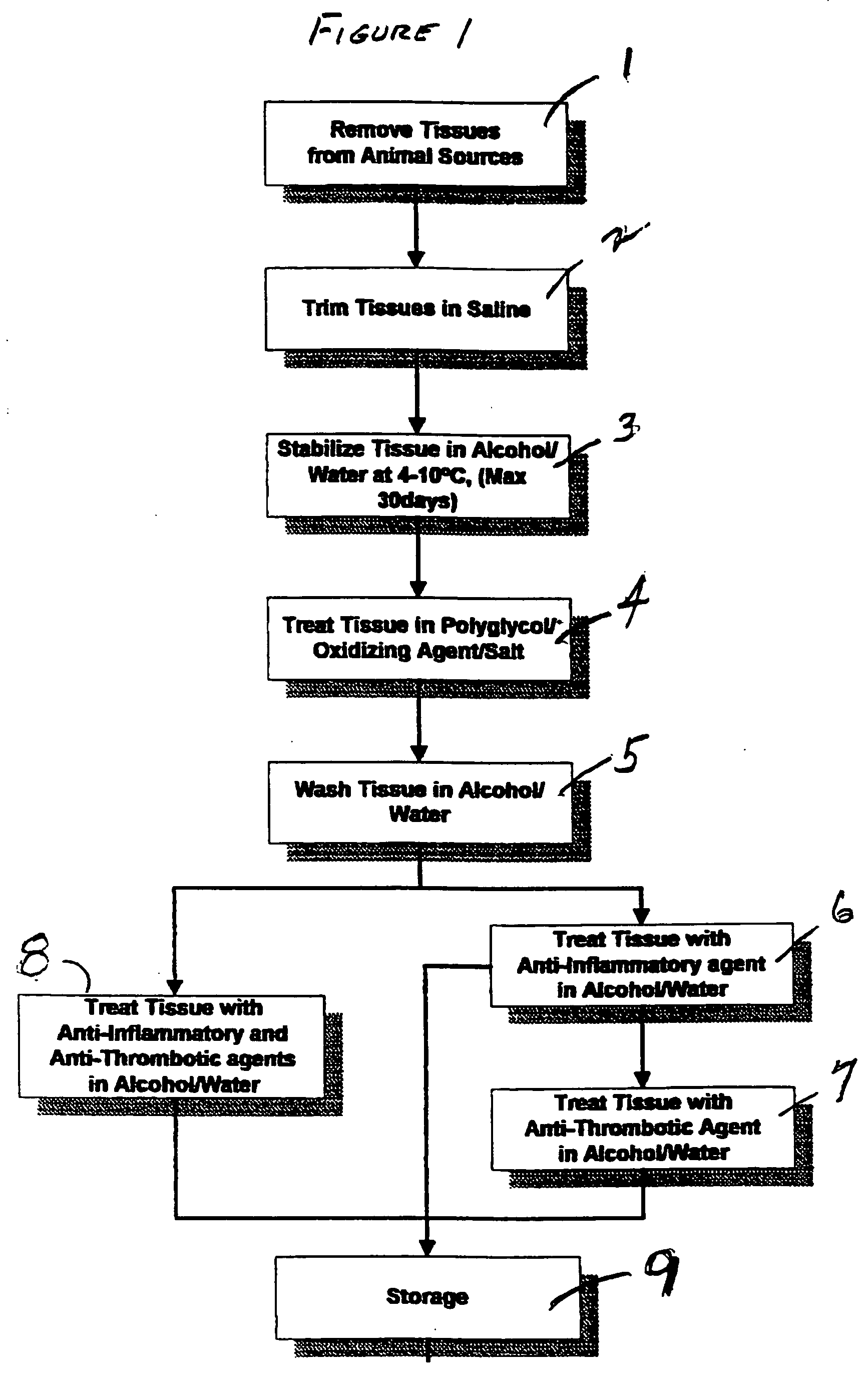
[0042] Freshly harvested bovine mesenteric arteries were stored in 50% alcohol for one week and then treated (or soaked) in a solution containing 5% PEG (molecular weight 8,000 D), 0.5% hydrogen peroxide, 4 M NaCl and 0.05 M phosphate buffered at pH 7.0 for 96 hours at 4° C. The arteries were washed in 50% ethanol twice and then further treated (or soaked) in a solution of indomethacin (50 mg / l) and heparin (250 IU / ml) in 50% ethanol overnight. The treated arteries were further soaked in 50% ethanol containing 20% glycerol, 5% PEG (MW=10,000), indomethacin (50 mg / l), and heparin (2501 U / ml). The arteries were then freeze-dried using a FTS Durastop / Duratop lyophilyzer system. The freeze-dried arteries were subjected to hydrogen peroxide sterilization by Star Service (Hayward, Calif.) with a Sterrad-100 hydrogen peroxide gas phase sterilizer using a standard validated sterilization program. The tissues were rinsed in saline for 30 minutes before the following studies were done.

1. Sh...

example 2

[0048] Freshly harvested bovine pericardium was stored in 50% alcohol for one week and then treated in a solution containing 5% PEG, 0.5% hydrogen peroxide, 4 M NaCl and 0.05 M phosphate buffered at pH 7.0 for 48 hours at 4° C. The pericardial tissues were washed in 50% ethanol twice and then further treated in a solution of indomethacin (50 mg / l) and heparin (250 IU / ml) in 50% ethanol overnight. The treated pericardial tissues were further soaked in 50% ethanol containing 20% glycerol, 5% PEG (MW=10,000), indomethacin (50 mg / l), and heparin (2501 U / ml). The pericardial tissues were then freeze-dried using a FTS Durastop / Duratop lyophilyzer system. The freeze-dried pericardial tissues were subjected to hydrogen peroxide sterilization by Star Service (Hayward, Calif.) with a Sterrad-100 hydrogen peroxide gas phase sterilizer using a standard validated sterilization program. The tissues were rinsed in saline for 30 minutes before the following study was done:

[0049] 1. Cell Adhesion a...

example 3

[0051] Bovine mesenteric arteries and porcine mammary arteries were stored in 50% alcohol for one week and then treated in a solution containing 5% PEG, 0.5% hydrogen peroxide, 4 M NaCl and 0.05 M phoshate buffered at pH 7.0 for 48 hours at 4° C. The arteries were washed in 50% ethanol twice and then further treated in a solution of indomethacin (20 mg / l) and heparin (250 IU / ml) in 50% ethanol overnight. The tissues were rinsed in saline for 30 minutes before the following studies were done:

[0052] 1. Sheep descending aorta to circumflex coronary artery bypass implant study—treated porcine mammary arteries, glutaraldehyde treated porcine and bovine conduits as well as PTFE grafts were implanted in 10 sheep as descending aorta to circumflex artery bypass for up to 202 days. The results of this study are summarized in the Table 3 below:

TABLE 3AnimalGroupPO Days*Patency1Treated Porcine Artery202SPatent2Treated Porcine Artery194SPatent3Treated Porcine Artery162SPatent4Treated Porcine ...


PUM

PropertyMeasurementUnit
temperatureaaaaaaaaaa
concentrationaaaaaaaaaa
molecular weightaaaaaaaaaa

Abstract

The invention relates to a method of treatment of collagenous connective tissue removed from a donor for implant into a recipient which is re-habited or re-colonized by host cells without an immune rejection and inflammatory reaction. After removal from the donor, the tissue is trimmed and thereafter soaked in a cold stabilizing solution having a temperature range of 4 to 10 degrees centigrade. The tissue is then soaked at a predetermined temperature in a polyglycol, salt, hydrogen peroxide, and phosphate buffer first solution of predetermined quantities and concentrations and of sufficient ionic strength to permit ground substances to dissociate such that the collagen fibers remain stable. The tissue is then soaked in an alcohol and water solution at a predetermined temperature for a sufficient period of time to remove the residue of the first solution. Following the removal of the residue, the tissue is soaked at a predetermined temperature in a third solution of an anti-inflammatory agent, an anti-thrombic agent, alcohol, and water or sequentially in an anti-inflammatory agent, alcohol, and water solution, and then in an anti-thrombic agent, alcohol and water solution and thereafter stored.

Description

[0001] This Application is a Divisional Application of Prior application Ser. No. 10 / 235,017, filed Sep. 23, 2002.FIELD OF THE INVENTION [0002] The present invention relates to a process for treating collagenous connective tissue from an animal donor for implant in an animal or human recipient. BACKGROUND OF THE INVENTION [0003] The present invention relates to a process for treating collagenous connective tissue or structural support tissue removed from an animal donor for implant into a recipient without an immune and inflammatory rejection. Collagenous connective tissue or structural support tissue may be heart valve tissue, blood vessel, pericardium, omentum, fascia, tendon, ligament, intestine, cartilage, bone, membrane, or other such tissue. [0004] Implants from animal donors into a human recipient to correct defective body components are known in the prior art. For instance, hemostatic collagen implants and collagen injections have been used for hemostasis and tissue augmenta...

Claims


Application Information

Patent Timeline
Patent Type & Authority Applications(United States)
IPC IPC(8): A61K35/34A61K38/39A61L27/36C12N5/08
CPCA01N1/02A01N1/0226A61K38/39A61L27/3604A61L27/3687
Inventor CHEUNG, DAVID T.
Owner CHEUNG DAVID T
Why Eureka
Try Eureka