Protein fragment complementation assays for high-throughput and high-content screening

a protein fragment and high-content technology, applied in the field of protein fragment complementation assays for high-throughput and high-content screening, can solve the problems of inefficiency of the current process, inability to detect new drugs, and inability to meet the needs of patients, so as to accelerate the identification of potential therapeutic compounds

Inactive Publication Date: 2006-10-05
ODYSSEY THERA INC
View PDF9 Cites 18 Cited by
  • Summary
  • Abstract
  • Description
  • Claims
  • Application Information

AI Technical Summary

Benefits of technology

[0132] Construction of an HTS or HCS assay An overview of the process of constructing an assay for HTS or HCS is shown in FIG. 1. The genes to be used in the HTS or HCS assay may code either for known or for novel interacting proteins. The interacting proteins can be selected by one or methods that include bait-versus-library screening; pairwise (gene by gene) interaction mapping; and / or prior knowledge of a pathway or an interacting protein pair. In the diagram, proteins numbered 3 and 4 are known (or can be shown) to participate in a receptor-mediated cell signaling pathway and can be chosen to construct an HTS or HCS screen to identify compounds that block the pathway. It should be noted that not all protein-protein complexes will be responsive to agonists, antagonists, activators or inhibitors of pathways. Some interactions will be constitutive. It is an advantage of the present invention that PCA can be used to identify protein-protein pairs that serve as ‘sentinels’ capable of reporting out the activity of a pathway. In either case, once the genes of interest are identified, the assays are constructed according to the following scheme: A reporter fragment pair F1 / F2 is generated (a partial list of reporters is in Table 1). Using for example two genes of interest encoding the protein-protein pair denoted as (3,4) in FIG. 1, two expression constructs are made, one comprising gene ‘3’ fused in frame to a flexible linker and to the F1 reporter fragment, and the other comprising gene ‘4’ fused in frame to a flexible linker and to the F2 reporter fragment, in such a way that the gene of interest, linker and reporter fragment are in frame and are operably linked to a promoter. (Polycistronic vectors can also be used. A complete description of vector options is given in Example 12). In FIG. 1 the genes are fused at the 5′ end and the encoded proteins of interest will be at the N-terminus of the fusions; other combinations, and details of vector construction and vector elements, are shown in FIG. 16. Cells are co-transfected with complementary F1, F2 gene constructs such that proteins are expressed. Transient assays can be performed; also, stable cell lines can be constructed with “PCA Inside” by using selectable markers or by using a survival selection PCA to generate the stable cell line. The resulting cells or stable cell lines are used for HTS or HCS in conjunction with chemical libraries of interest.
that PCA can be used to identify protein-protein pairs that serve as ‘sentinels’ capable of reporting out the activity of a pathway. In either case, once the genes of interest are identified, the assays are constructed according to the following scheme: A reporter fragment pair F1 / F2 is generated (a partial list of reporters is in Table 1). Using for example two genes of interest encoding the protein-protein pair denoted as (3,4) in FIG. 1, two expression constructs are made, one comprising gene ‘3’ fused in frame to a flexible linker and to the F1 reporter fragment, and the other comprising gene ‘4’ fused in frame to a flexible linker and to the F2 reporter fragment, in such a way that the gene of interest, linker and reporter fragment are in frame and are operably linked to a promoter. (Polycistronic vectors can also be used. A complete description of vector options is given in

Problems solved by technology

Pharmaceutical company investment in new drug discovery and development has increased dramatically over the last ten years, yet the rate of new drug approvals has not kept pace.
Expensive pre-clinical and clinical failures are responsible for much of the inefficiency of the current process.
Although such assays are inexpensive to perform, they can take 6 months or longer to develop.
A major problem is that the development of an in vitro assay requires specific reagents for every target of interest, including purified protein for the target against which the screen is to be run.
Often it is difficult to express the protein of interest and / or to obtain a sufficient quantity of the protein in pure form.
Moreover, although in vitro assays are the gold standard for pharmacology and studies of structure activity relationships, in vitro screening does not provide information about the biological availability or activity of the compound hit.
Although the optical instrumentation and hardware have advanced to the point that any bioluminescent signal can be detected with high sensitivity and high throughput, the existing assay choices are limited either with respect to their range of application, format, biological relevance, or ease of use.
Although bait-versus-library screening with Y2H has been carried out in high throughput, several features of Y2H limit its utility for functional protein target validation and for screening of chemical libraries.
First, Y2H often requires the expression of the proteins of interest within the nucleus of a cell such as the yeast cell, which is an unnatural context for most human proteins and cannot be used at all for human membrane proteins such as receptors.
Second, yeast do not contain the human biochemical pathways that are of interest for drug discovery, which obviates pathway-based discovery and validation of novel, potential drug target proteins.
Third, except for chemicals that directly disrupt protein-protein interactions, Y2H is not of use in identifying pharmacologically active molecules that disrupt mammalian biochemical pathways.
Quantifying FRET or BRET-can be technically challenging and use in imaging protein-protein interactions is very limited due to the very weak FRET signal.
However, at the present time FRET and BRET are not easily amenable to high-throughput screening of either cDNA libraries or chemical libraries as we describe below.
However, the adaptation of beta-gal complementation to live cell assays has been limited because the phenomenon occurs naturally, resulting in significant background activity.
On the other hand, beta-gal is not suitable for high-content assays because the product of the beta-gal reaction diffuses throughout the cell.
A PCA fragment is not a luminophore and does not enable monitoring of the redistribution of an individual protein.

Method used


Image

  • Protein fragment complementation assays for high-throughput and high-content screening
  • Protein fragment complementation assays for high-throughput and high-content screening
  • Protein fragment complementation assays for high-throughput and high-content screening

Examples


example 1

Fluorescent and Luminescent Assays for HTS and HCS

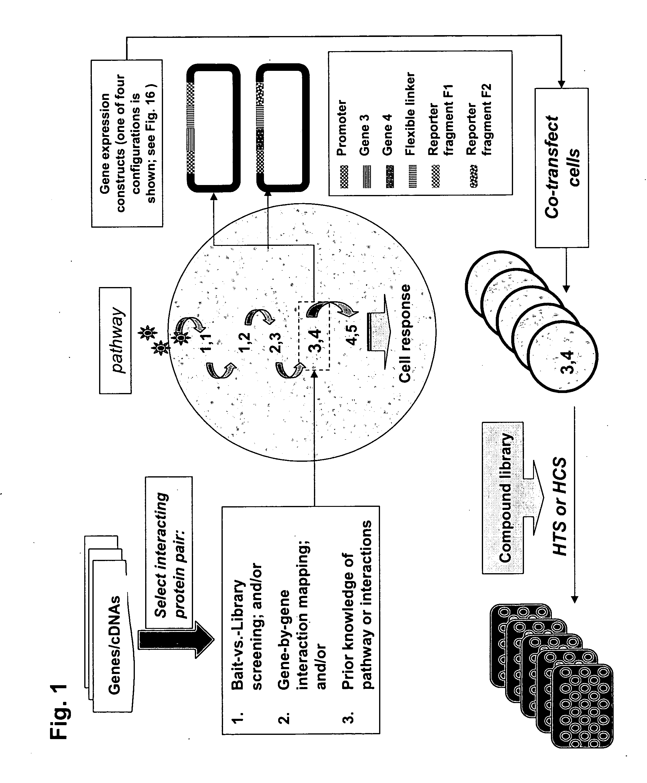
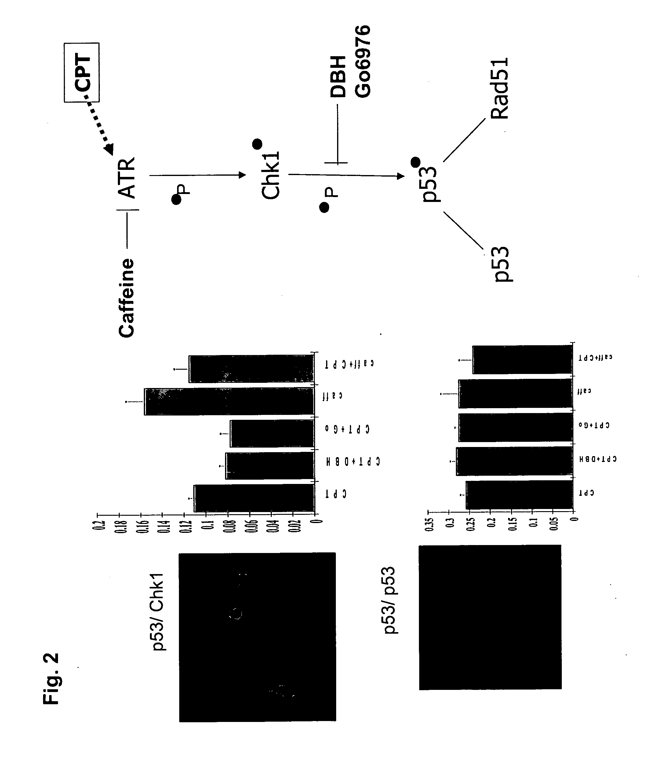
[0142] In the first example of the present invention, we sought to demonstrate the construction of a wide range of useful assays based on fluorescent and luminescent PCAs and to demonstrate their use for high-throughput and high-content assays in conjunction with standard HTS and HCS instrumentation. We used elements of the DNA damage response pathway.

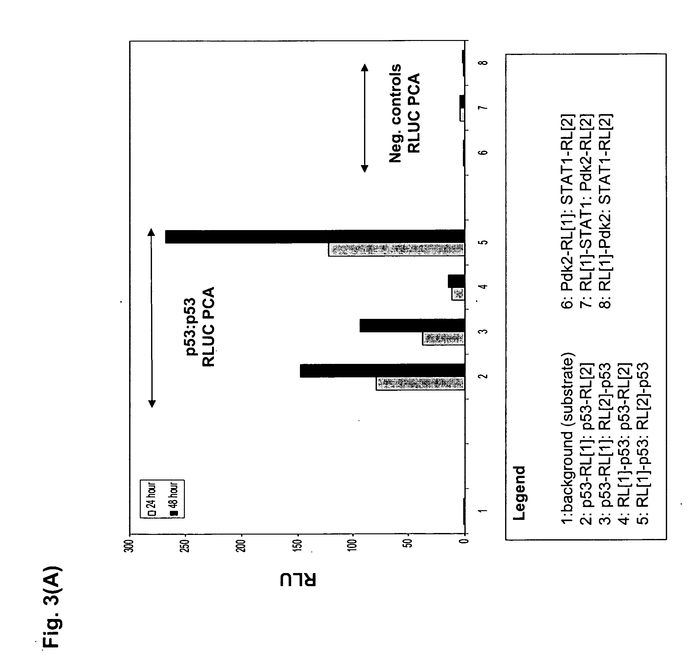
[0143]FIG. 2 shows a scheme of the pathway and the results for an HTS assay based on a beta-lactamase PCA (BLA PCA) and a HCS assay based on a GFP PCA. FIG. 3 shows an HTS assay based on Renilla luciferase (RLuc PCA). FIG. 4 shows a high-content assay based on an IFP PCA (IFP is a variant of GFP). In these examples, the proteins assayed are interacting pairs in the DNA damage response pathway, specifically, the checkpoint kinase Chk1 which interacts with the tumor suppressor p53 (Chk1 / p53 PCA), or p53 itself which forms homodimers (p53 / p53 PCA).

[0144] For the BLA PCA, the genes of in...

example 2

Identifying Novel Protein-Protein Interactions and Constructing Quantitative Assays

[0166] The PCA strategy described in the invention and depicted in FIG. 1 was next used to identify novel protein-protein interactions in the PI-3-kinase and PKA / PKC-mediated pathways and then to carry out quantitative screens based on the novel interactions. First, cDNA library screening was performed with the GFP PCA in order to identify proteins interacting with PKB. A novel interaction between PKB and Ft1 was identified by the GFP PCA screen. Subsequently, the GFP PCA was used to construct fluorescent assays for PKB / Ft1 and PDK1 / Ft1. The organization of the pathways and the position of the PKB / hFt1 interaction is shown in FIG. 5(A). Methods for library screening with PCA and for assay construction are provided below.

[0167] DNA Constructs. The full-length cDNAs encoding PKB, PDK1, PKCalpha, the catalytic subunit of PKA (PKAc), GSK3beta, BAD, Caspase 9 and FKHRL1 were amplified by PCR and subclone...

example 3

High-Throughput Assays with YFP PCA

[0171] We next sought to demonstrate that PCAs can be used as quantitative assays providing relevant pharmacological information. For the example we used the well-characterized interaction of FKBP (the FK506 binding protein) and its cognate partner, FRAP (FKBP-Rapamycin-Associated-Protein), an interaction that occurs only at a low level in untreated cells but which is markedly induced by the immunosuppressant drug, rapamycin. The organization of the human growth pathway, showing the ‘sentinel’ FKBP / FRAP interaction, is depicted in FIG. 6A.

[0172] For these studies, we used a YFP PCA. HEK 293E Cells were seeded into a 96 well plates at a cell density of 13,000 per well. Cell media is MEM-alpha Growth medium. Total volume was 100 microliters. Cells were allowed to grow 20-24 hours prior to transfection—cells were 70-80% confluent at time of transfection. Cells were maintained at 37C, 5% CO2. Cells were transfected with a total of 0.1 micrograms of D...


PUM

PropertyMeasurementUnit
timeaaaaaaaaaa
volumeaaaaaaaaaa
concentrationaaaaaaaaaa

Abstract

The present invention provides protein fragment complementation assays for drug discovery, in particular to identify compounds that activate or inhibit cellular pathways. Based on the selection of an interacting protein pair combined with an appropriate PCA reporter, the assays may be run in high-throughput or high-content mode and may be used in automated screening of libraries of compounds. The interacting pair may be selected by cDNA library screening; by gene-by-gene interaction mapping; or by prior knowledge of a pathway. Fluorescent and luminescent assays can be constructed using the methods provided herein. The selection of suitable PCA reporters for high-throughput or high-content (high-context) assay formats is described for a diversity of reporters, with particular detail provided for examples of monomeric enzymes and fluorescent proteins. Methods are described for constructing such assays for one or more steps in a biochemical pathway; testing the effects of compounds from combinatorial, natural product, peptide, antibody, nucleic acid or other diverse libraries on the protein or pathway(s) of interest; and using the results of the screening to identify specific compounds that activate or inhibit the protein or pathway(s) of interest. Single-color and multi-color assays are disclosed. Further disclosed are universal expression vectors with cassettes that allow the rapid construction of assays for a large and diverse number of gene / reporter combinations. The development of such assays is shown to be straightforward, providing for a broad, flexible and biologically relevant platform for drug discovery.

Description

[0001] This application claims the priority benefit under 35 U.S.C. section 119 of U.S. Provisional Patent Application No. 60 / 445,225 entitled “Protein fragment complementation assays for high-throughput and high-content screening”, filed Feb. 6, 2003, which is in its entirety herein incorporated by reference. This Application is also a continuation-in-part of pending U.S. application Ser. No. 10 / 353,090 filed Jan. 29, 2003; which application is a continuation of pending U.S. application Ser. No. 10 / 154,758 filed May 24, 2002; which is a continuation of U.S. Ser. No. 09 / 499,464 filed Feb. 7, 2000; and now U.S. Pat. No. 6,428,951; which is a continuation of U.S. Ser. No. 09 / 017,412 filed Feb. 2, 1998; and now U.S. Pat. No. 6,270,964. The entire contents of all those patents and applications are incorporated by reference herein.BACKGROUND OF THE INVENTION [0002] Pharmaceutical company investment in new drug discovery and development has increased dramatically over the last ten years, ...

Claims


Application Information

Patent Timeline
Patent Type & Authority Applications(United States)
IPC IPC(8): C40B30/02G06F19/00C07K14/00C12N1/21C12N15/11C12Q1/00C12Q1/68G01NG01N33/50G01N33/53G01N33/543G01N33/567
CPCG01N33/5008G01N33/5011G01N2500/02G01N33/542G01N33/6845G01N33/5041Y02A90/10
Inventor WATSON MICHNICK, STEPHENREMY, INGRIDMACDONALD, MARNIELAMERDIN, JANEYU, HELENWESTWICK, JOHN
Owner ODYSSEY THERA INC
Why Eureka
Try Eureka