System and method for integrating ancillary data in DICOM image files

Active Publication Date: 2006-10-26
MEDIMAGING TOOLS
View PDF36 Cites 78 Cited by
  • Summary
  • Abstract
  • Description
  • Claims
  • Application Information

AI Technical Summary

Benefits of technology

[0013] A system and method is further provided that enables a user to easily select from a list of patients, and that converts files from a first format, such as the Joint Photographic Experts Group (“JPEG”) format, to a DICOM format using DICOM tags and data of the selected patient's imaging examination to “marry” this information to the imaging exam

Problems solved by technology

Teleradiologists (i.e., physicians who read and/or interpret medical images at a remote location) have encountered a major obstacle regarding images formatted according to the DICOM standard.
Unfortunately, such information is not typically available and teleradiologists often encounter an obstacle because they cannot interpret patient examinations accurately in a vacuum.
However, in practice, such information is often not provided or used as intended.
This occurs, typically, because technologists who are time-constrained, tend to omit data entry tasks that they perceive as overly time-consuming, and they rationalize that the radiologist who eventually reads and interprets the medical image(s) already has or can gain access easily to the patient's chart should the radiologist require more in

Method used


Image

  • System and method for integrating ancillary data in DICOM image files
  • System and method for integrating ancillary data in DICOM image files
  • System and method for integrating ancillary data in DICOM image files

Examples


Example

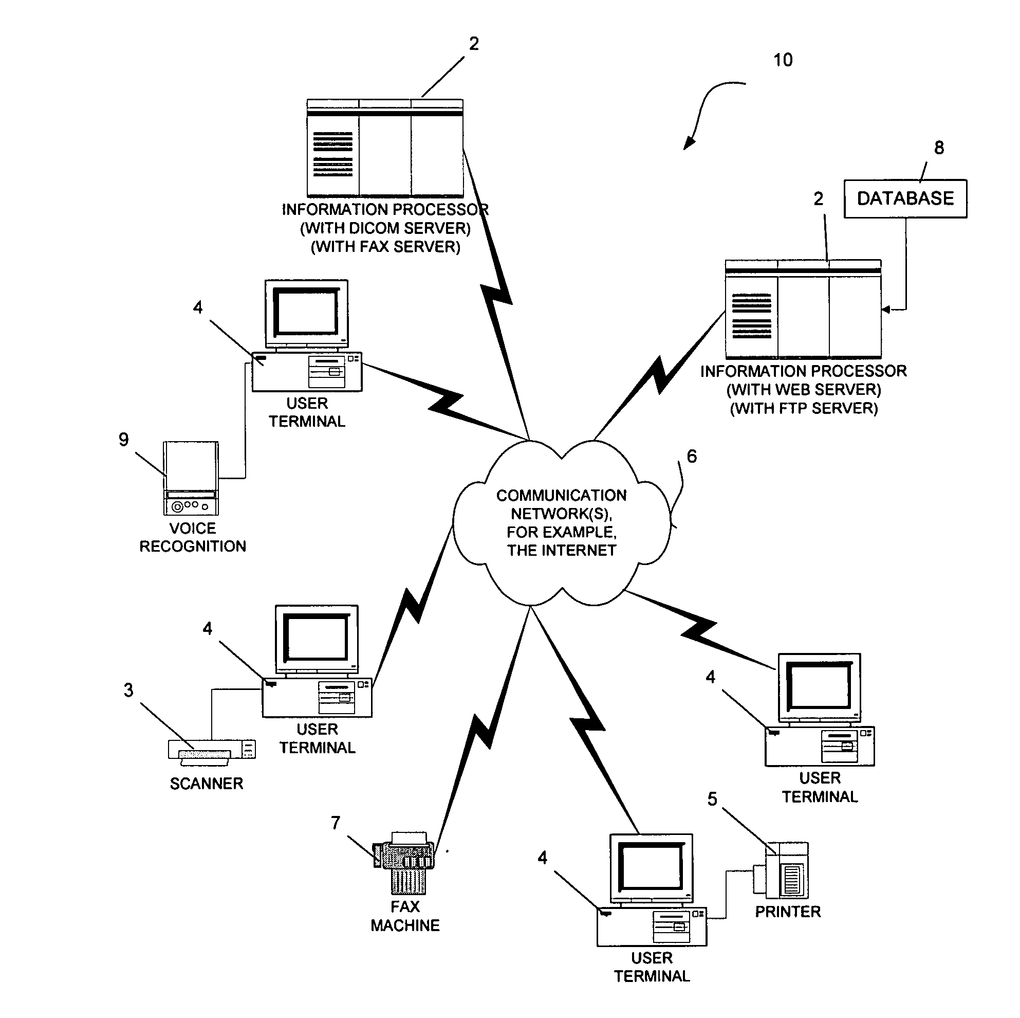
[0030] The present invention comprises a system and method for incorporating paperwork / handwritten (analog) clinical patient information into digital DICOM image files for interpretation by the radiologist or other physician. In an example embodiment, the present invention comprises a suite of software modules which reads and incorporates paperwork / handwritten (analog) clinical patient information directly integrating them into the DICOM image examination files.

[0031] In an example embodiment of the present invention, patient demographic information is output as a separate file after a patient examination file is acquired by a DICOM server. This output file comprised of key available DICOM tags, contains demographic information of the patient along with detailed information regarding the examination itself. A key indexing number, for example, labeled the “Study UID” number, is preferably included among these DICOM tags in the output file. The present invention imports the output fi...


PUM


Abstract

The invention comprises a system and method for integrating electronic patient information with an electronic medical image file. In one embodiment, at least one information processor is provided that is operable to communicate over a communication network. A database is provided on the at least one information processor, wherein the database is operable to store electronic patient information representing a plurality of patients, and further to store respective image files representing patient medical examinations. A receiving module is preferably included that receives an image file representing a patient medical examination. The image file is preferably formatted with a respective filename. Also, a converting module is provided that is operable to convert the image file to be associated with at least one tag, wherein the converting module refers to at least the respective filename to associate the image with the tag(s), and further the tag(s) are associated with the image file to associate the medical image record with the electronic patient information for a respective patient. A storage module is further operable to store the converted image to the database. Preferably, the modules operate substantially without human intervention.

Description

CROSS-REFERENCE TO RELATED APPLICATIONS [0001] The present application is based on and claims priority to U.S. Provisional Patent Application Ser. No. 60 / 655,331, filed on Feb. 22, 2005 and entitled SYSTEM AND METHOD FOR INTEGRATING ANCILLARY DATA IN DICOM IMAGE FILES, U.S. Provisional Patent Application Ser. No. 60 / 661,773, filed on Mar. 15, 2005 and entitled SYSTEM AND METHOD FOR INTEGRATING ANCILLARY DATA IN DICOM IMAGE FILES (II),” and U.S. Provisional Patent Application Ser. No. 60 / 670,326, filed on Apr. 12, 2005 and entitled “SYSTEM AND METHOD FOR INTEGRATING ANCILLARY DATA IN DICOM IMAGE FILES (III),” the entire contents of all of which are hereby incorporated by reference.BACKGROUND OF THE INVENTION [0002] 1. Field of the Invention [0003] The present invention relates, generally, to medical image records, and, more particularly, with providing DICOM image files with medical information received from at least one or more sources. [0004] 2. Description of the Related Art [0005...

Claims


Application Information

Patent Timeline
IPC IPC(8): G06F7/00
CPCG06F17/30265G06Q50/24G06Q50/22G06F19/321G06F16/58G16H30/20G16H40/67
Inventor ROTHPEARL, ALLENKILLCOMMONS, JOHN
Owner MEDIMAGING TOOLS
Why Eureka
Try Eureka