Registration of three dimensional image data to 2D-image-derived data

a three-dimensional image and data technology, applied in image analysis, image enhancement, instruments, etc., can solve the problem of difficult for a physician to become oriented in a three-dimensional setting using a single-plane x-ray image projection display

Active Publication Date: 2006-11-30
STEREOTAXIS
View PDF93 Cites 49 Cited by
  • Summary
  • Abstract
  • Description
  • Claims
  • Application Information

AI Technical Summary

Benefits of technology

[0006] Another embodiment of the present invention may further provide a method for automatically determining a centerline for the corresponding three-dimensional image data of the vessel using a software algorithm, determining a direction of the vessel from the two points obtained from the X-ray image planes, and calculating a transformation of the centerline direction derived from the three-dimensional image data to the direction derived from the X-ray image data points to obtain a registration of the three-dimensional image data with the two-dimensional image data of the X-ray imaging system.
[0007] Another embodiment of the present invention may provide a method for registration of three-dimensional anatomical image data with two-dimensional X-ray imaging system data to allow for overlay of pre-operative anatomical images onto the X-ray imaging planes.

Problems solved by technology

In some cases, it may be difficult for a physician to become oriented in a three dimensional setting using a display of a single-plane X-ray image projection.

Method used


Image

  • Registration of three dimensional image data to 2D-image-derived data
  • Registration of three dimensional image data to 2D-image-derived data
  • Registration of three dimensional image data to 2D-image-derived data

Examples


second embodiment

[0018] In a second embodiment, the software could reconstruct the 3D vessel centerline and display it as a 3D model, possibly as a tubular surface, in a 3D window on a Graphical User Interface. The user could select pairs of points as described above, directly on the 3D model, and these could be used for the registration. In this case the 2D images are not directly used to perform the registration with the 3D anatomical data; instead the 3D reconstruction derived from the 2D images is.

third embodiment

[0019] In a third embodiment, the user marks at least 4 non-coplanar branch points in the 2D-image-derived data (which could either be directly the 2D images, or a 3D reconstruction derived from 2D images, as described above), and corresponding branch points in the 3D anatomical data. The software then performs a landmark registration between the data sets using the branch points as landmark points, using a standard method such as the Procrustes algorithm.

fourth embodiment

[0020] In a fourth embodiment, the user could mark a set of branch points and at least one direction corresponding to a vessel take-off orientation at a branch, and the software performs a best-fit registration based on finding a rigid transformation that minimizes a weighted sum of cost functions based on distances and orientations respectively.

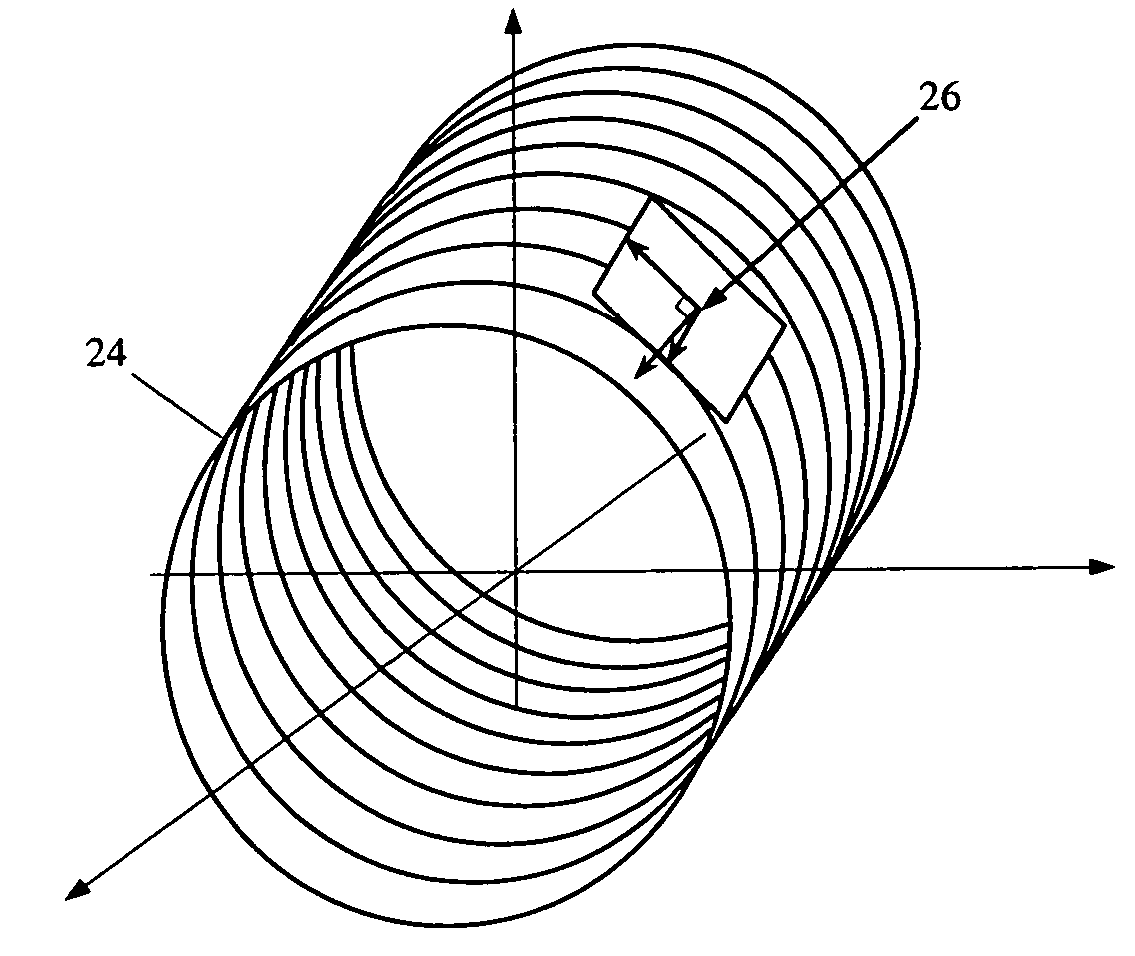
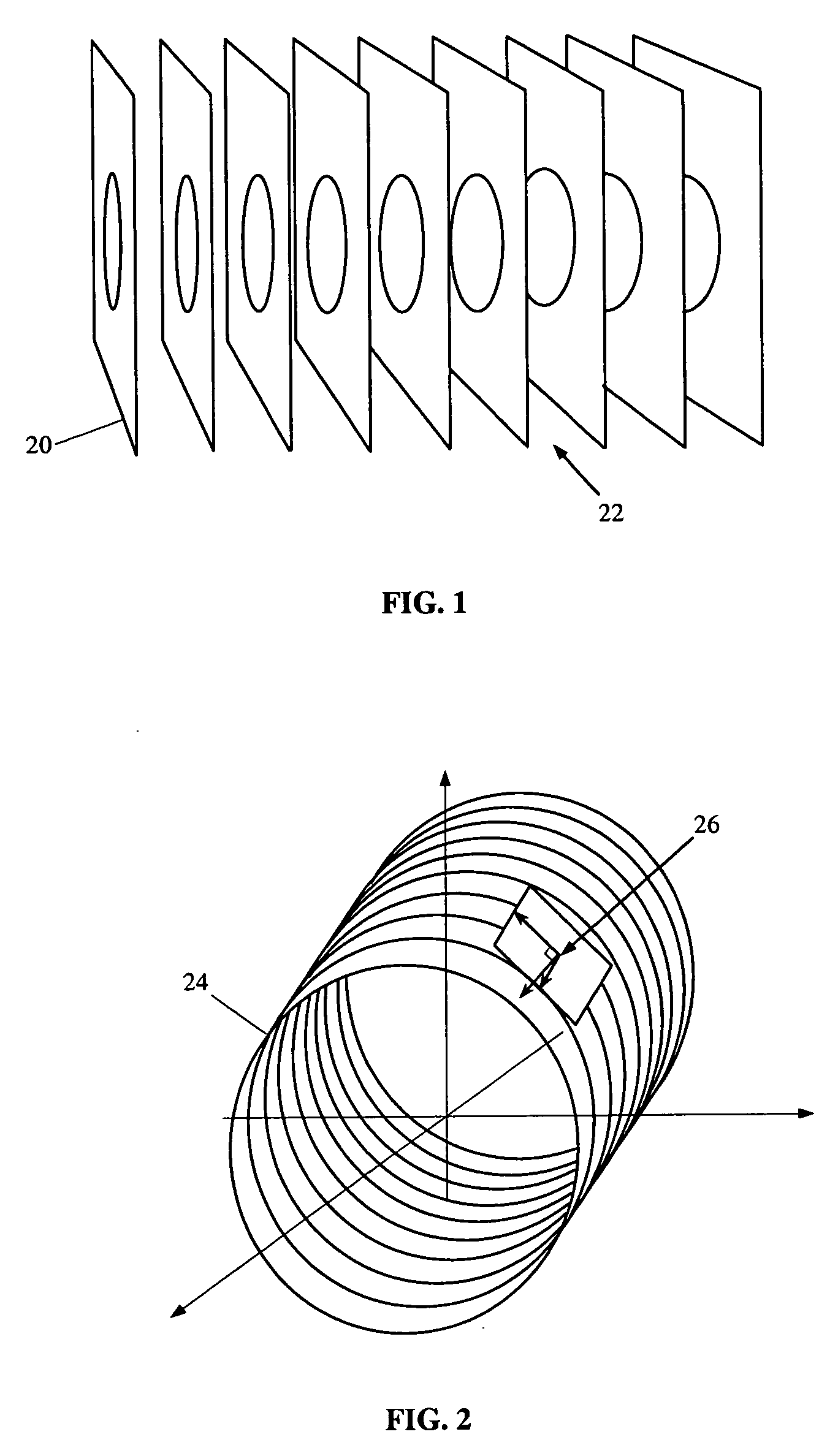
[0021] The method of the various embodiments initially calls for the user to mark on at least two X-ray image planes two points on an anatomical vessel that provide a linear indication of the local direction of the vessel, which two anatomical vessel points have coordinates y1 and y2. The method then analyzes a three-dimensional image such as shown in FIG. 1 having a series of cross-sectional images 20 of a volume that was regularly sampled at some constant interval to create an image pixel data set that is utilized by image modeling software. The spaces 22 between each section are filled in so that the pixel data take on additional dimensio...


PUM


Abstract

A method for obtaining registration of a three-dimensional image data set of an anatomical vessel with corresponding two-dimensional image data of the vessel in an X-ray imaging system, where the method comprise the user identifying two points on an anatomical vessel on at least two X-ray image planes, the user identifying two similar points on the surface of the corresponding three-dimensional anatomical image data, determining the orientation direction of the vessel from the two user identified image data surface points, determining the orientation direction of the vessel from the two points obtained from the X-ray image planes, and calculating a transformation of the three-dimensional image data to obtain a best fit registration of the direction derived from the image surface points with the direction derived from the X-ray image data points.

Description

CROSS-REFERENCE TO RELATED APPLICATIONS [0001] This application claims the benefit of U.S. Patent Application Ser. No. 60 / 650,616, filed Feb. 7, 2005, the entire disclosure of which is incorporated herein by reference.FIELD OF THE INVENTION [0002] This invention relates to registration of three-dimensional data to a reference coordinate system, and more particularly to registration of three dimensional image data with an X-ray imaging coordinate system. BACKGROUND OF THE INVENTION [0003] Interventional medicine is the collection of medical procedures in which access to the site of treatment is made by navigation through one of the subject's blood vessels, body cavities or lumens. Interventional medicine technologies have been applied to manipulation of medical instruments which contact tissues during surgical navigation procedures, making these procedures more precise, repeatable and less dependent of the device manipulation skills of the physician. Some presently available interven...

Claims


Application Information

Patent Timeline
Patent Type & Authority Applications(United States)
IPC IPC(8): G06K9/00
CPCG06K9/00208G06T7/0028G06T7/0038G06T2207/30101G06T2207/10121G06T2207/30048G06T2207/10072G06T7/33G06T7/38G06V20/647
Inventor VISWANATHAN, RAJU R.
Owner STEREOTAXIS
Why Eureka
Try Eureka