Beam therapy treatment user interface monitoring and recording system

a technology of user interface and recording system, applied in the field of beam therapy treatment user interface monitoring and recording system, can solve problems such as failure to provide electronic digital recording

Active Publication Date: 2006-12-14
CERNER INNOVATION
View PDF3 Cites 18 Cited by
  • Summary
  • Abstract
  • Description
  • Claims
  • Application Information

AI Technical Summary

Problems solved by technology

These diagrammatic representations or photographed hand marked anatomy images are time consuming to produce, potentially inaccurate and fail to provide an electronic digital record.

Method used


Image

  • Beam therapy treatment user interface monitoring and recording system
  • Beam therapy treatment user interface monitoring and recording system
  • Beam therapy treatment user interface monitoring and recording system

Examples


Embodiment Construction

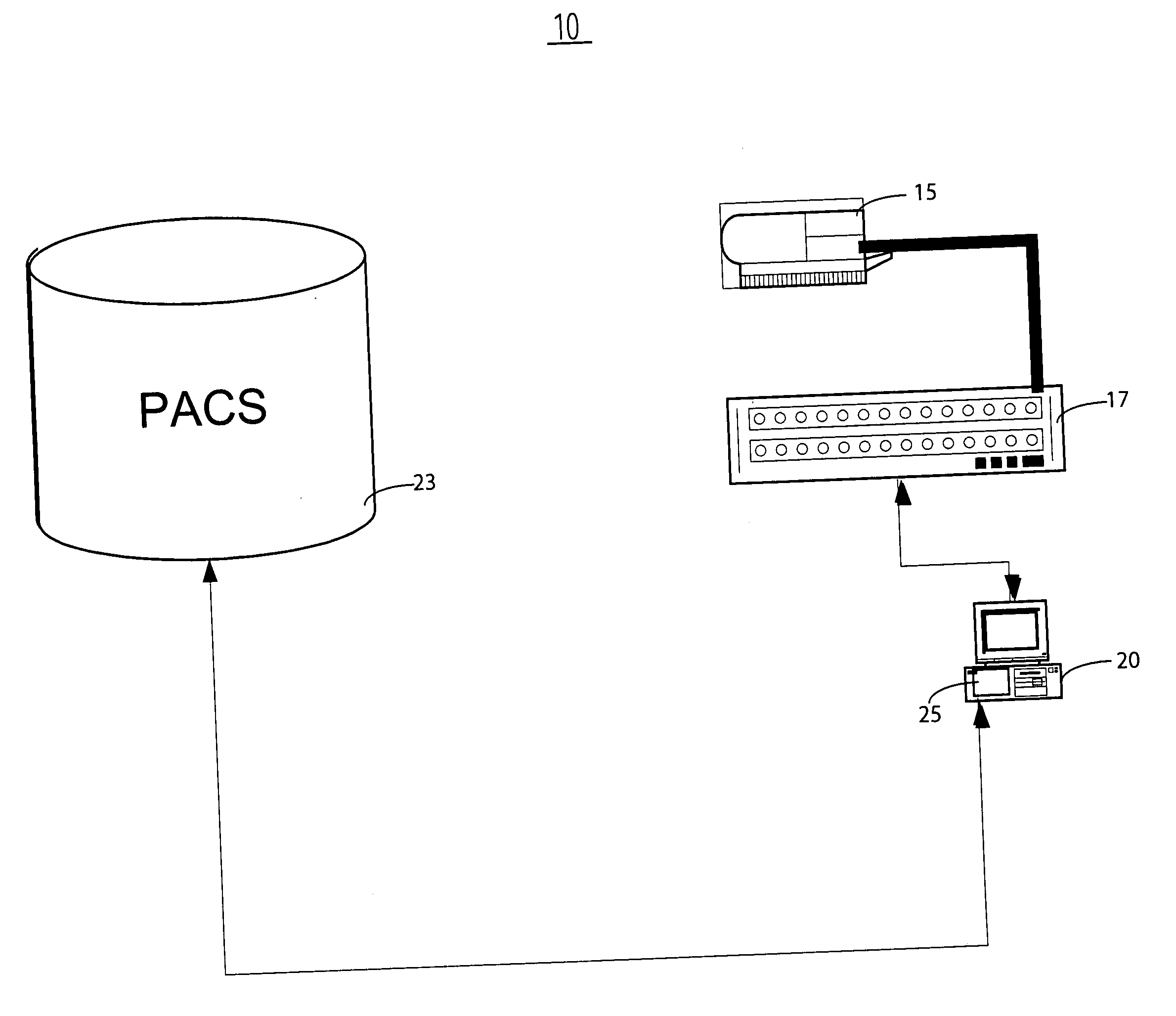
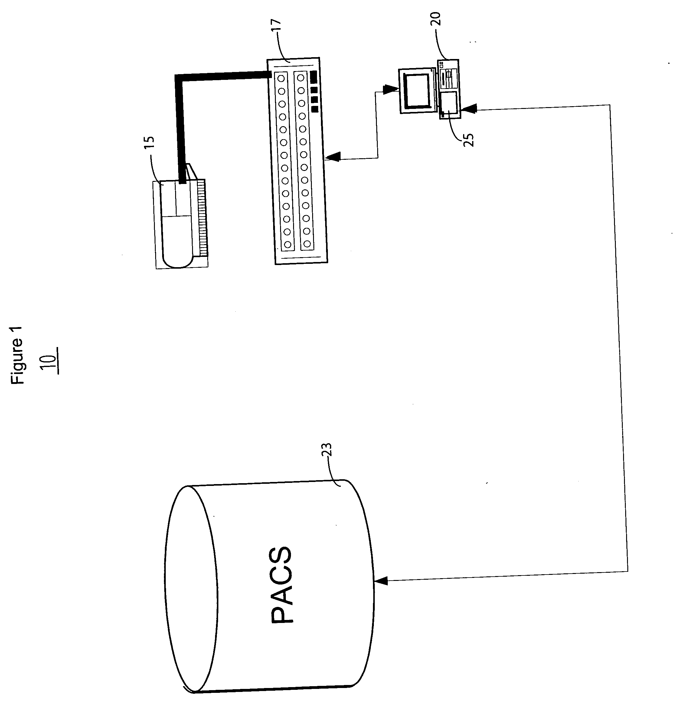
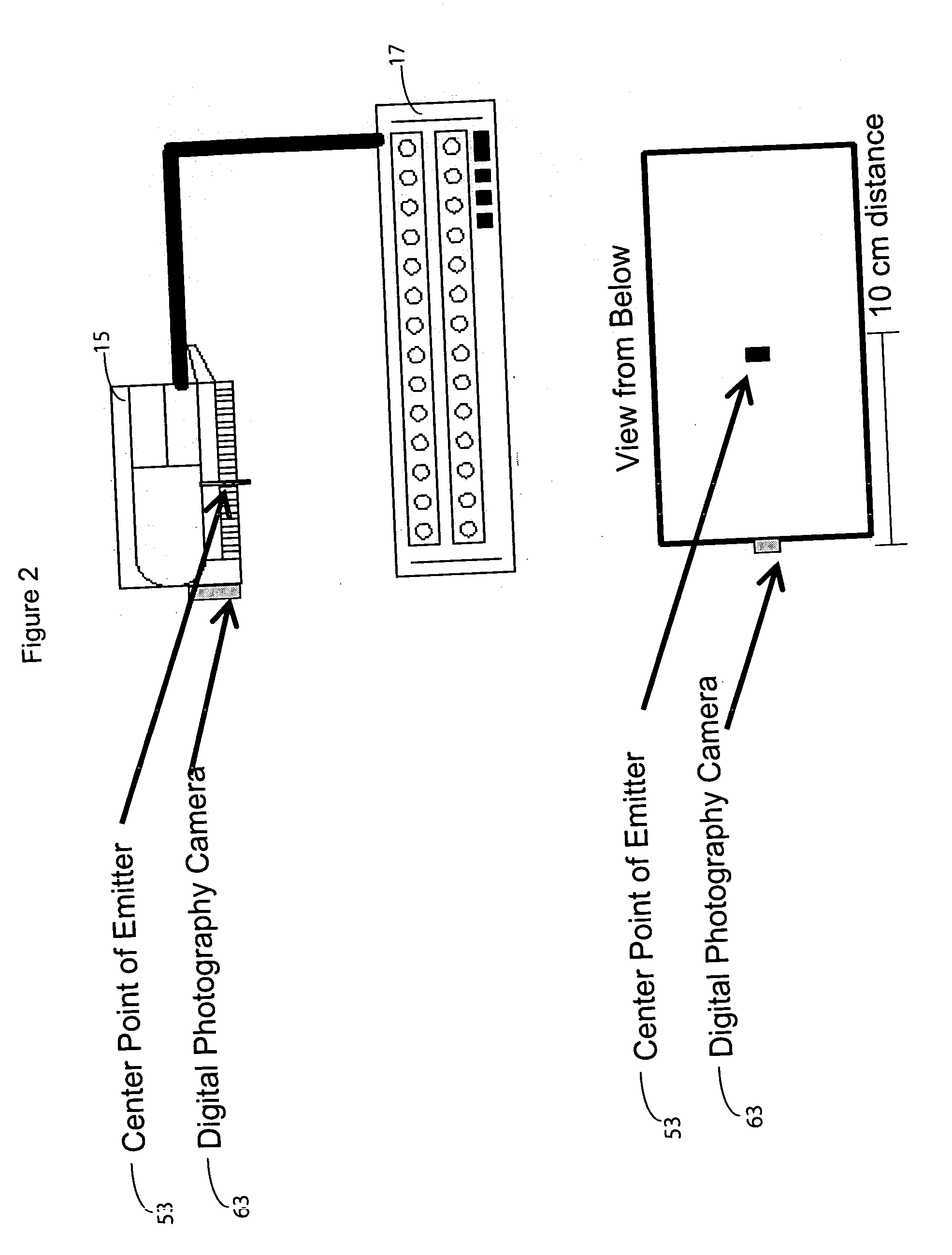
[0012] A system enables physicists and radiation oncology personnel to accurately store information regarding positioning of a radiation beam and its contact points on the surface of, and within, the anatomy of a patient. A radiation oncologist needs accurate data regarding previous radiation dosage received by a region of patient anatomy in order to determine a current dosage and treatment of a patient and to ensure no single area of tissue is overexposed to radiation. This information is needed in a timely fashion to support seamless oncologist workflow (task sequence) performance. The system enables radiation oncology personnel to accurately view a radiation beam (two dimensional and three dimensional) contact region within a patient without use of subjective, potentially inaccurate drawings. The system employs a digital photographic camera attached to a radiation oncology therapy device used in treating cancer to create an image overlay providing information indicating radiation...


PUM


Abstract

A system uses a digital photographic camera attached to a radiation therapy device to create an overlay of radiation beam information comprising data identifying treated patient anatomical region data and supporting determination of cumulative radiation treatment information from multiple treatment overlays. A system records a treatment beam landing area on the anatomical body surface of a patient for use in radiation or other therapy. The system includes an input processor for receiving data representing an image of the surface of a portion of the anatomy of a patient. A data processor provides beam landing position data relative to the image by calculating a position of a treatment beam landing area relative to the image using received input data indicating projection characteristics of a treatment beam used in treating the portion of the anatomy of the patient. A storage processor stores the beam landing position data in a record associated with the patient.

Description

[0001] This is a non-provisional application of provisional application Ser. No. 60 / 690,119 by M. P. Esham et al. filed Jun. 13, 2005.FIELD OF THE INVENTION [0002] This invention concerns a system for the recording of a treatment beam landing area on the anatomical body surface of a patient for use in radiation or other therapy. BACKGROUND OF THE INVENTION [0003] Healthcare workers in a radiology department of a hospital, for example, need to record and document locations (area and volume) on patient anatomy where radiation beam therapy occurs. In existing radiology departments, radiation personnel typically document beam placement using representation diagrams. The radiation personnel also (or alternatively) directly mark patient skin and photograph the marked areas. These diagrammatic representations or photographed hand marked anatomy images are time consuming to produce, potentially inaccurate and fail to provide an electronic digital record. A system according to invention prin...

Claims


Application Information

Patent Timeline
Patent Type & Authority Applications(United States)
IPC IPC(8): A61N5/10
CPCA61N2005/1054A61N5/1049
Inventor ESHAM, MATTHEW PAULFITZGERALD, LORETTA A.CHI, ANDREW
Owner CERNER INNOVATION
Why Eureka
Try Eureka