Image analysis method for vertebral compression curvature

Inactive Publication Date: 2007-02-15
CHUNG YUAN CHRISTIAN UNIVERSITY
View PDF5 Cites 4 Cited by
  • Summary
  • Abstract
  • Description
  • Claims
  • Application Information

AI Technical Summary

Benefits of technology

[0008] Since the B-spline curve has a good ability in approximating circles and arcs, the disclosed method can thus clo

Problems solved by technology

The diagnosis method for vertebral compression curvature, no matter from clinical findings or image diagnosis such as X-ray films, computed tomography (CT), and magnetic resonance imaging (MRI), cannot very accurately find out what the real problems are.
Normal image diagnosis methods cannot p

Method used


Image

  • Image analysis method for vertebral compression curvature
  • Image analysis method for vertebral compression curvature
  • Image analysis method for vertebral compression curvature

Examples


Embodiment Construction

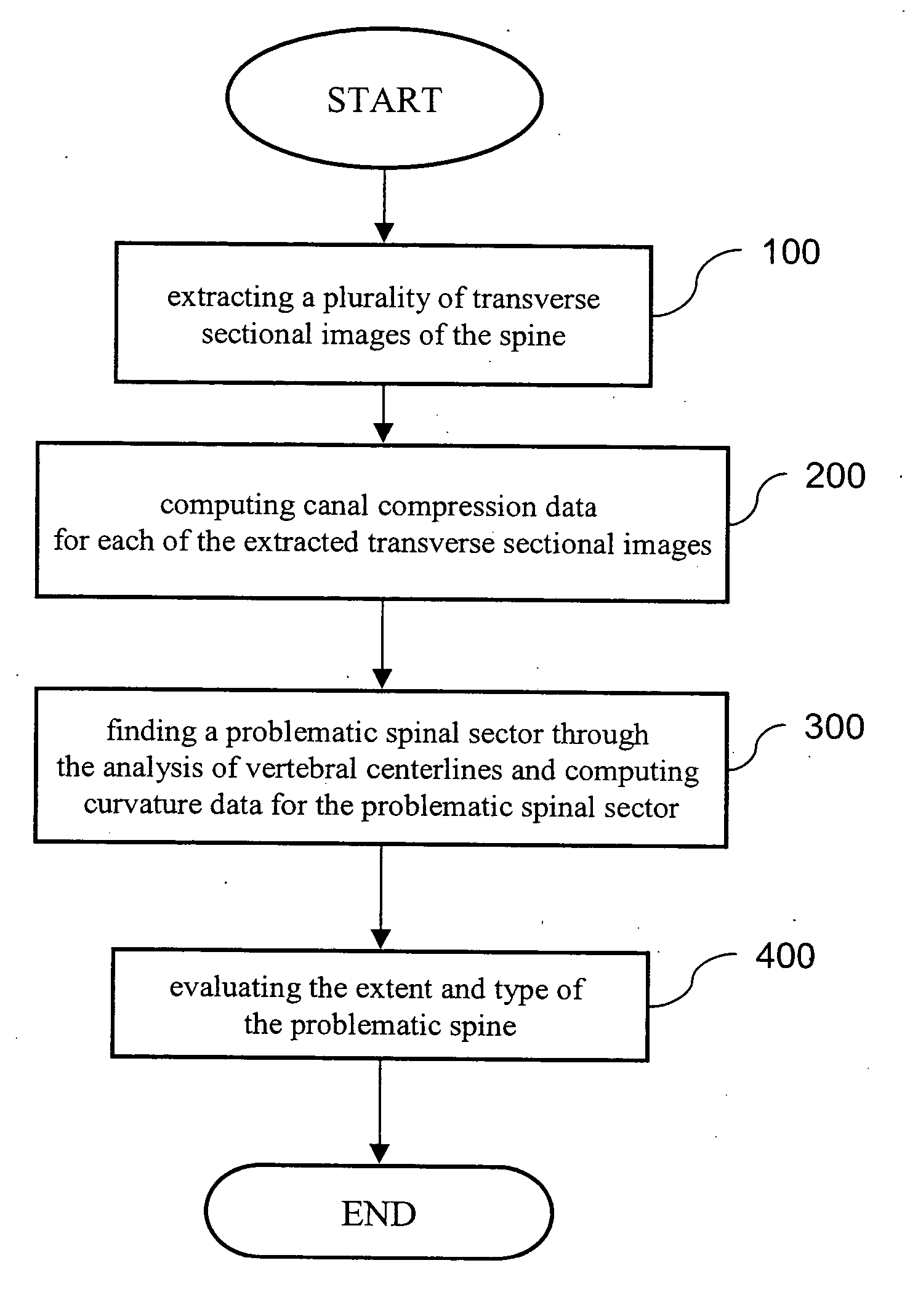
[0022] The invention discloses an image analysis method for vertebral compression curvature. It is primary used to perform diagnostic analysis of the vertebral compression curvature caused by pressure or fracture. First, we use FIG. 1 to explain the main procedure of the disclosed method.

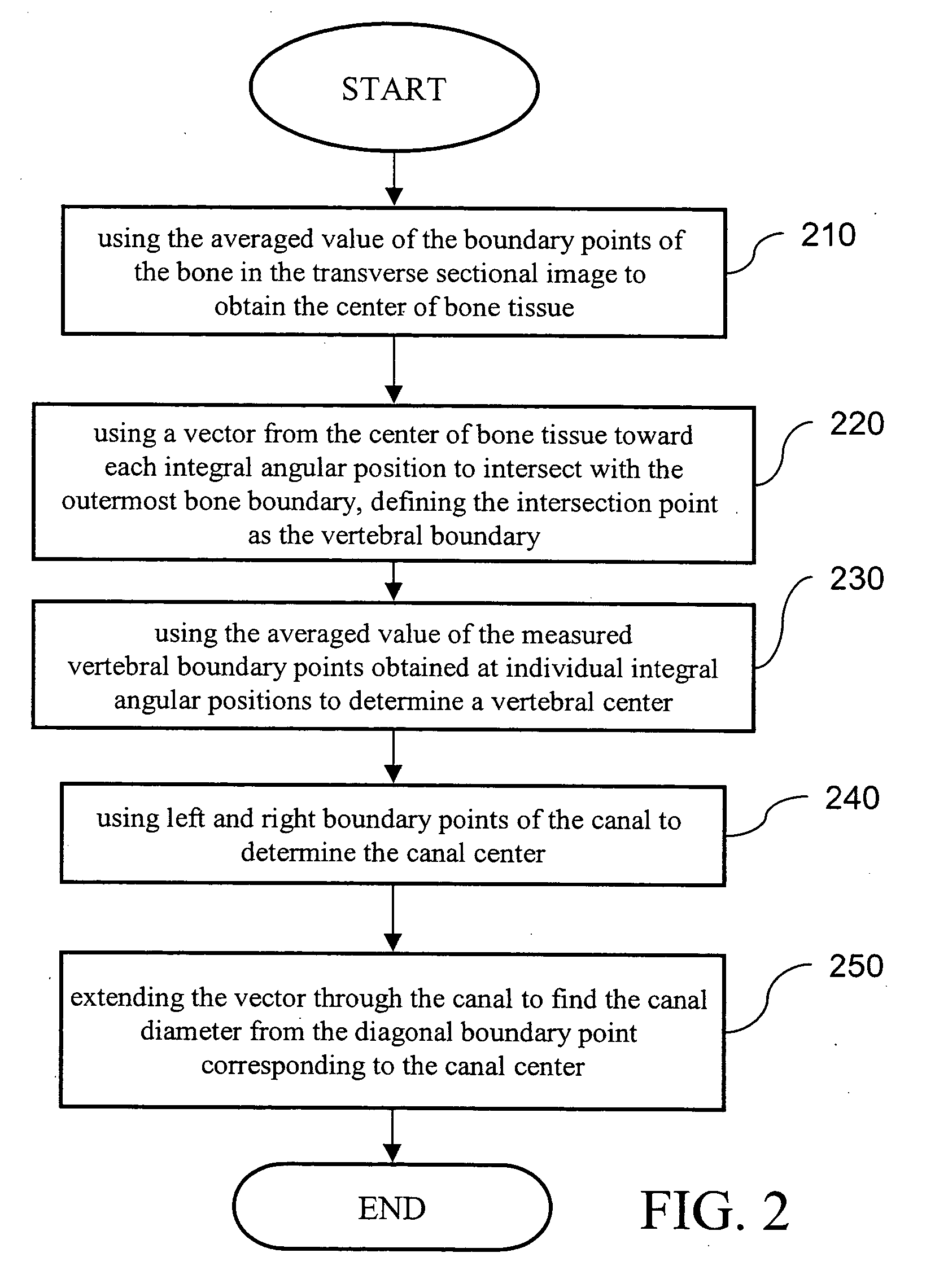
[0023] In the beginning, we use computed tomography (CT) or magnetic resonance imaging (MRI) to extract transverse sectional images of the spine to be analyzed (step 100). In general, the extraction location, extraction spacing, and extraction amount are different as the results obtained from preliminary X-ray films vary. Each transverse sectional image is computed to obtain the compression data of the canal in it (step 200). Such data include the canal diameter, the three-dimensional coordinates of the canal center, and so on. This is because the canal diagonal part of the spine is most likely to be depressed by external forces and to be deformed. Therefore, the method uses this principle to compa...


PUM


Abstract

An image analysis method for vertebral compression curvature is disclosed for providing diagnosis analysis of the compression curvature. It makes use of the transverse sectional image with a concave feature of a vertebral body. After B-spline curves are approximated as ellipse-like surfaces, the method further evaluates the compression curvature of the canal. On the other hand, the center of the ellipse-like surface boundary obtained by approximation from different transverse sectional images of the vertebral body is used to reconstruct the centerline of the vertebral body by linear restoration. Such information is used to determine the curvature of the vertebral body. Moreover, the method can use the above-mentioned reconstructed vertebral body centerline to compare with other adjacent vertebral centerlines that have normal curvatures. In this manner, the method can help determine the type and extent of the spine under pressure or having a fracture.

Description

BACKGROUND OF THE INVENTION [0001] 1. Field of Invention [0002] The invention relates to an image analysis method and, in particular, to a method that diagnoses the spine compression curvature from the transverse sectional image of the spine. [0003] 2. Related Art [0004] The diagnosis of spine compression curvature and particularly in determining the extent and type of the compression curvature has been the hardest part in medical sciences. However, the diagnosis information in this respect is the most valuable part in surgical operations and / or therapeutic procedures. [0005] The diagnosis method for vertebral compression curvature, no matter from clinical findings or image diagnosis such as X-ray films, computed tomography (CT), and magnetic resonance imaging (MRI), cannot very accurately find out what the real problems are. The main reason is that the most accurate diagnosis method has to be companied with the three-dimensional image analysis for abnormal spines and the analysis b...

Claims


Application Information

Patent Timeline
IPC IPC(8): G06K9/00A61B5/05G06T5/00G06T7/60
CPCG06T7/0012G06T7/604G06T2207/30012G06T2207/10088G06T2207/20044G06T2207/10081G06T7/64
Inventor TSAI, MING-DARHSIEH, MING-SHIUM
Owner CHUNG YUAN CHRISTIAN UNIVERSITY
Why Eureka
Try Eureka