
Tissue cutting devices having hemostasis capability and related methods

a cutting device and tissue technology, applied in the field of medical devices, can solve the problems of difficult diagnosis of tissue samples, and achieve the effect of effectively stopping the bleeding at the tissue si

Inactive Publication Date: 2007-03-22
BOSTON SCI SCIMED INC
View PDF15 Cites 14 Cited by
  • Summary
  • Abstract
  • Description
  • Claims
  • Application Information

AI Technical Summary

Benefits of technology

[0004] Therefore, various exemplary embodiments of the invention may provide biopsy forceps having a separate hemostatic device integrated with the biopsy forceps, which may effectively stop the bleeding at the tissue site without damaging the tissue samples obtained by the biopsy forceps.

Problems solved by technology

Such “hot” biopsy forceps, however, may damage the tissue samples collected in the biopsy forceps, such that a proper diagnosis of the tissue samples may be difficult.

Method used


Image

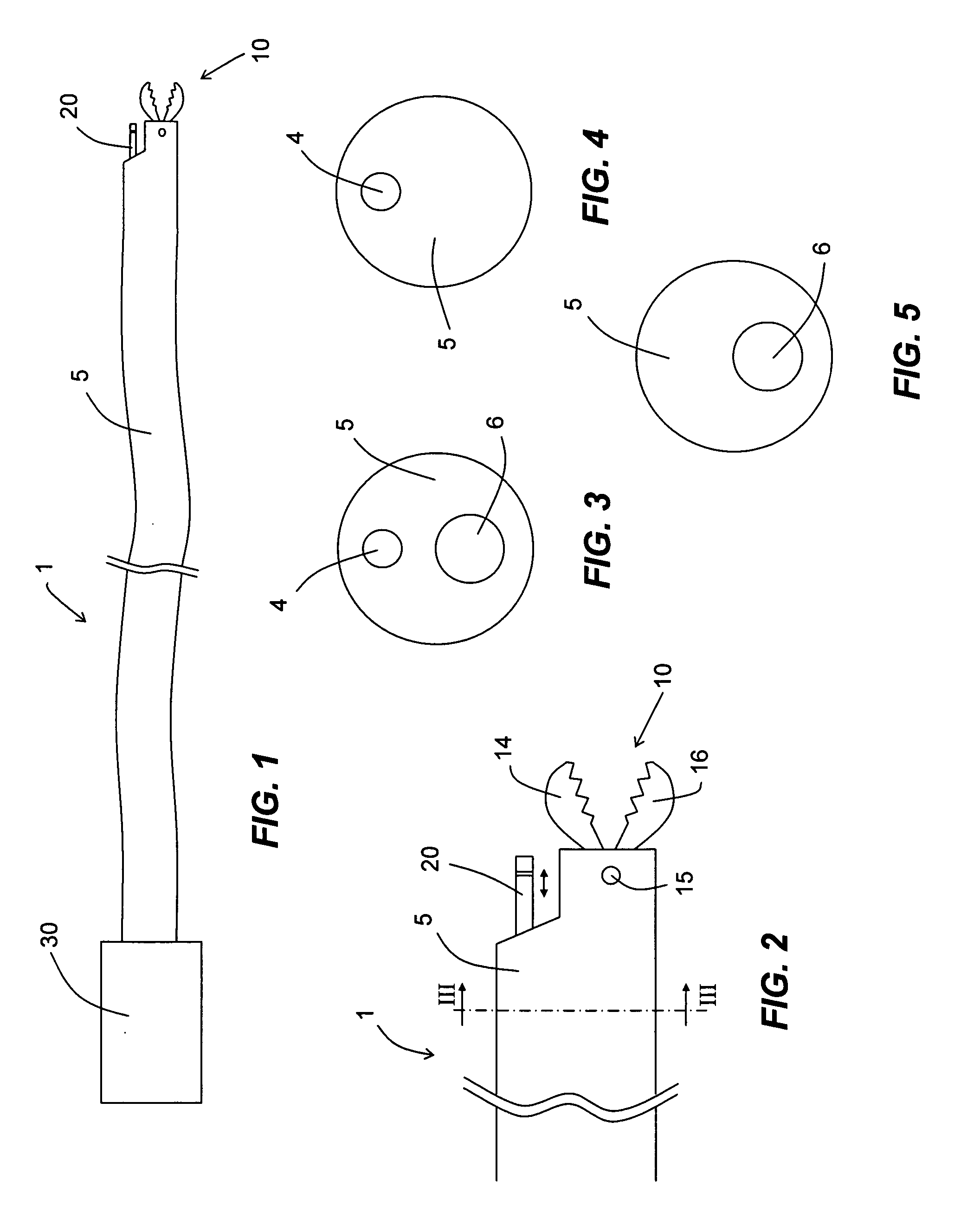
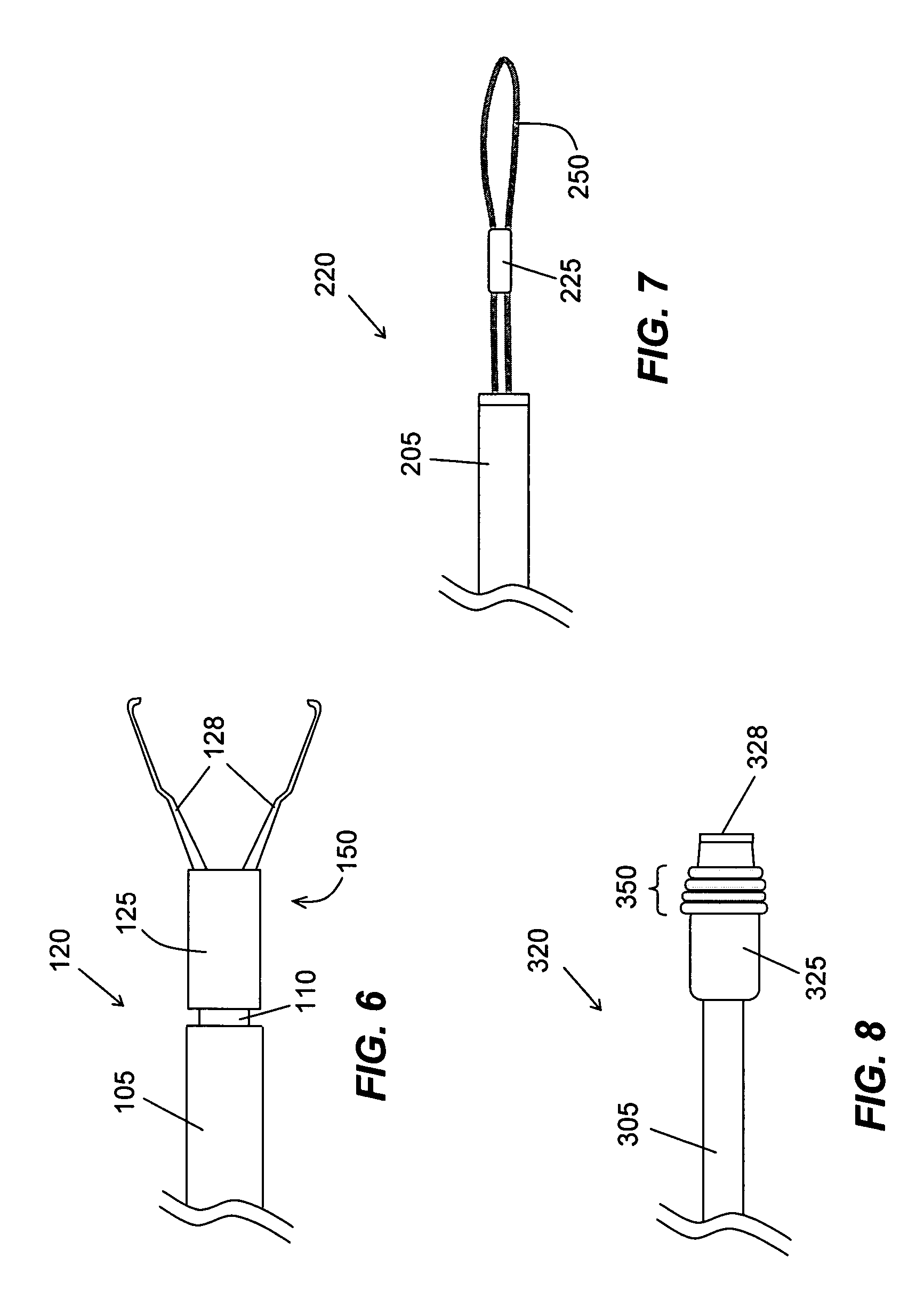
  • Tissue cutting devices having hemostasis capability and related methods
  • Tissue cutting devices having hemostasis capability and related methods
  • Tissue cutting devices having hemostasis capability and related methods

Examples


Embodiment Construction

[0021] Reference will now be made in detail to the exemplary embodiments consistent with the present invention, examples of which are illustrated in the accompanying drawings. Wherever possible, the same reference numbers will be used throughout the drawings to refer to the same or like parts.

[0022]FIG. 1 illustrates an endoscopic medical device 1 (e.g., tissue acquisition device) having a separate hemostatic device for use in, for example, obtaining tissue samples from a patient's body, according to an exemplary embodiment of the invention. While the invention will be described in connection with a particular tissue acquisition device (i.e., biopsy forceps), embodiments of the invention may be used with any other types of tissue acquisition device (e.g., snares, scissors, needles, pincers, knives, knifeneedles, or baskets). Moreover, certain aspects of the inventions may be applied to, or used in connection with, other numerous surgical applications involving tissue cutting that m...


PUM


Abstract

Various embodiments of a medical device (e.g., tissue acquisition device) having a separate hemostasis capability and related methods of use are disclosed. The device may include an elongated tubular member having a proximal end and a distal end, a tissue cutting member proximate the distal end of the tubular member, and a hemostatic member proximate the distal end of the tubular member and adjacent the tissue cutting member. At least one of the tissue cutting member and the hemostatic member may be fixedly connected to the tubular member.

Description

FIELD OF THE INVENTION [0001] The present invention relates generally to medical devices and related methods. More specifically, particular embodiments of the invention relate to tissue acquisition devices (e.g., biopsy forceps) having a separate hemostasis capability for use in, for example, acquiring tissue samples from a body. DESCRIPTION OF RELATED ART [0002] Biopsy forceps are widely used to obtain tissue samples. A biopsy forceps generally includes a distal cutting member configured to sever tissue samples and a tissue receptacle for holding one or more of the severed tissue samples. Severing tissue from a tissue site causes that tissue site to bleed. To stop or minimize the bleeding during the procedure, “hot” biopsy forceps may be used to direct cauterizing electric current or thermal energy to the tissue site through the distal cutting member to cauterize the bleeding tissue site. Such “hot” biopsy forceps, however, may damage the tissue samples collected in the biopsy forc...

Claims


Application Information

Patent Timeline
Patent Type & Authority Applications(United States)
IPC IPC(8): A61B17/32
CPCA61B10/06A61B17/10A61B17/3478A61B2017/22074A61B18/20A61B2017/12004A61B2017/12018A61B18/1492
Inventor GEITZ, KURT A.E.HOFFMAN, DAVID
Owner BOSTON SCI SCIMED INC
Learn More