Method and Apparatus for Standardizing Ultrasonography Training Using Image to Physical Space Registration of Tomographic Volumes From Tracked Ultrasound

a technology of ultrasonography and image to physical space, applied in the field of standardizing ultrasonography training, can solve the problems of irregular and unpredictable flow of patients, unpredictable duration of training, and difficulty in training students and technicians on even general ultrasonography

Inactive Publication Date: 2007-04-12
VANDERBILT UNIV
View PDF17 Cites 26 Cited by
  • Summary
  • Abstract
  • Description
  • Claims
  • Application Information

AI Technical Summary

Problems solved by technology

Such a system provides much more diagnostic information, but training students and technicians on even general ultrasonography is a somewhat complicated undertaking.
In a clinical setting, the flow of patients is irregular and unpredictable.
Therefore, the duration of training is unpredictable.
By using the time of a radiologist or other skilled imaging specialist along with the trainee, the time and cost expended is significant by the time the trainee is sufficie

Method used


Image

  • Method and Apparatus for Standardizing Ultrasonography Training Using Image to Physical Space Registration of Tomographic Volumes From Tracked Ultrasound
  • Method and Apparatus for Standardizing Ultrasonography Training Using Image to Physical Space Registration of Tomographic Volumes From Tracked Ultrasound
  • Method and Apparatus for Standardizing Ultrasonography Training Using Image to Physical Space Registration of Tomographic Volumes From Tracked Ultrasound

Examples


Embodiment Construction

[0027] Certain terminology is used in the following description for convenience only and is not limiting. The words “right”, “left”, “lower”, and “upper” designate directions in the drawings to which reference is made. The words “inwardly” and “outwardly” refer direction toward and away from, respectively, the geometric center of the object discussed and designated parts thereof. The terminology includes the words above specifically mentioned, derivatives thereof and words of similar import. Additionally, the word “a”, as used in the claims and in the corresponding portions of the specification, means “one” or “at least one.”

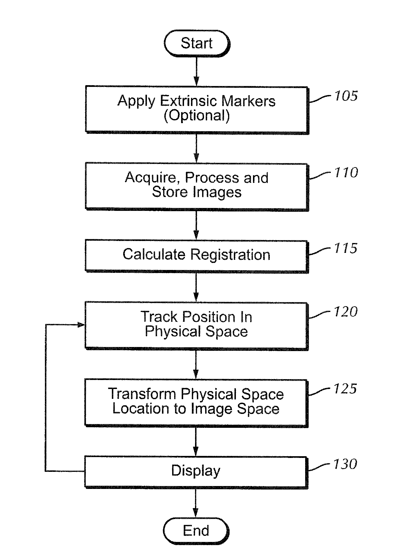
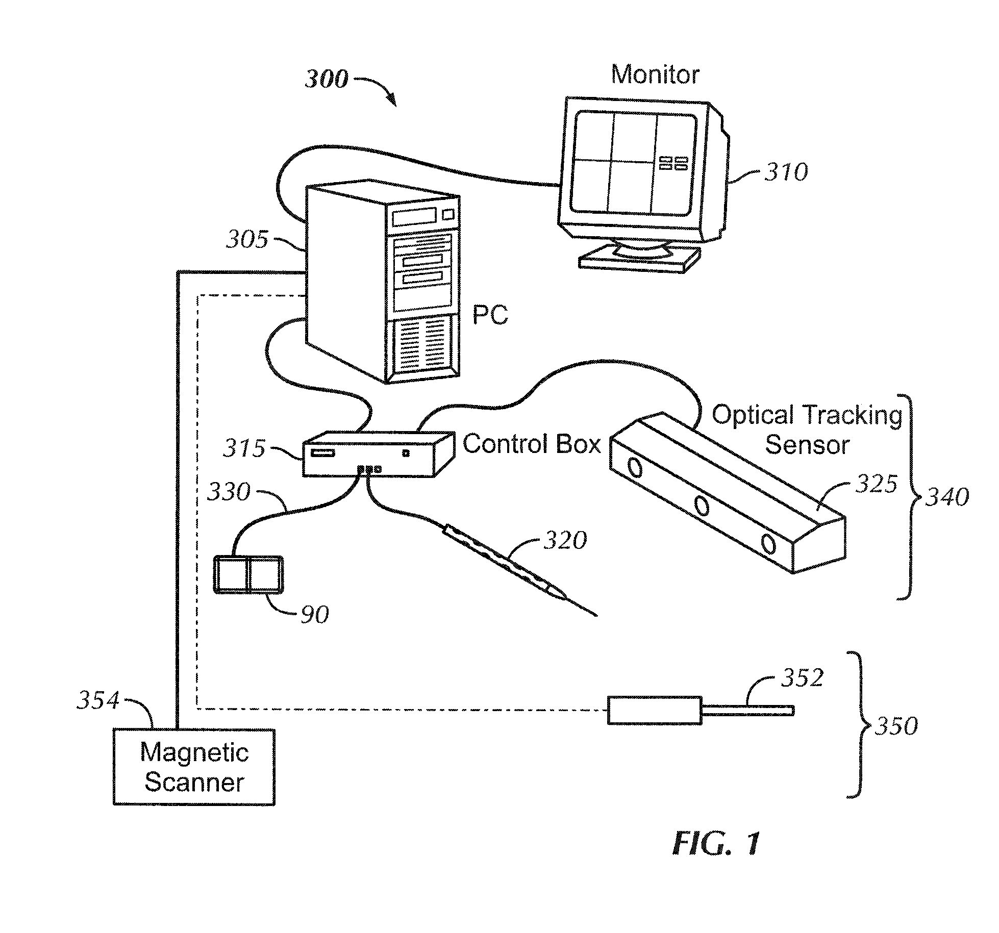
[0028] Preferred embodiments of the present invention include Image Guided Procedures (IGP). IGP have four basic components: image acquisition, image-to-physical-space registration, three-dimensional tracking, and display of imaging data and location. A relevant IGP system is disclosed in U.S. Pat. No. 6,584,339 B2 (Galloway, Jr. et al.), the contents of which i...


PUM


Abstract

A clinical and training apparatus that collects and processes physical space data while performing an image-guided procedure on an anatomical area of interest includes a calibration probe, a tracked ultrasonic probe, a wireless tracking device that tracks the ultrasonic probe in space and an image data processor. The physical space data provides three-dimensional coordinates for each of the physical points. The image data processor includes a memory holding instructions. The instructions include determining registrations used to indicate position in image space and physical space; using the registrations to map into image space, image data describing the physical space of the tracked ultrasonic probe and the anatomical area of interest; and constructing a three-dimensional (3D) volume based on ultrasonic image data. The apparatus includes a storage medium that stores a plurality of 3D volumes acquired by the image data processor for later retrieval.

Description

CROSS REFERENCE TO RELATED APPLICATIONS [0001] This application claims priority to U.S. Provisional Patent Application No. 60 / 720,813 filed on Sep. 27, 2005 entitled “Method and Apparatus for Standardizing Ultrasonography Training Using Image to Physical Space Registration of Tomographic Volumes from Tracked Ultrasound.”BACKGROUND OF THE INVENTION [0002] The present invention relates to a method and apparatus for standardizing ultrasonography training, and more particularly, a method and apparatus for standardizing ultrasonography training using image to physical space registration of tomographic volumes from tracked ultrasound. [0003] There are a number of medical imaging and instrumentation systems presently available including Positron Emission Tomography (PET), Magnetic Resonance Imaging (MRI), computed tomography (CT) and ultrasound. CT and MRI are imaging modalities that provide tomographic slices of the imaged volume. [0004] CT and / or MRI used in combination with other imagin...

Claims


Application Information

Patent Timeline
IPC IPC(8): G06K9/00
CPCG09B23/285A61B8/4245A61B8/4263
Inventor WARMATH, JOHN R.HERLINE, ALAN J.
Owner VANDERBILT UNIV
Why Eureka
Try Eureka