Apparatus and method for detecting deformability of cells using spatially modulated optical force microscopy

a spatial modulation and optical force technology, applied in the field of apparatus and method for detecting deformation of cells using spatial modulated optical force microscopy, can solve the problems of limited success, limitation of optical stretcher technique, and the inability to measure suspended cells

Inactive Publication Date: 2007-04-19
ARRYX INC
View PDF4 Cites 22 Cited by
  • Summary
  • Abstract
  • Description
  • Claims
  • Application Information

AI Technical Summary

Benefits of technology

[0013] Thus, with the present invention, optical deformability of any type of cell may be measured on the spatially modulated optical force microscope (SMOFM), so that diseased cells, including cancer cells, that have a unique optical deformability signature may be identified. In addition to being used as a diagnostic tool, the spatial light modulated force microscope may also be used as an investigative tool to understand the causes of any change in optical deformability due to a particular diseased cell state. Optical deformability of such cells may be correlated with structural protein expression levels and patterns, for example, allowing the spatially modulated force microscope to uncover the molecular origins for the changes in the viscoelastic responses of cells brought on by disease.
[0014] The apparatus of the present invention is optically based, so it has the advantages of being a quick and sterile measurement platform. In contrast to the prior art, the objects or cells to be probed in the present invention may be adhering to a surface (i.e., microscope cover slip surface), and so are able to retain their naturally present focal adhesions and stress fibers.
[0015] The present detection technique (using spatially modulated optical microscopy) is a much more sensitive technique for the detection of deformation, with sub-nanometer level sensitivity, compared to that of prior art methods. The greater sensitivity of the present invention has two important advantages: 1) a greater dynamic range of elastic responses may be probed, and 2) detectable deformations may be achieved at lower laser powers. The second advantage is an important consideration for parallelization.
[0017] In this way the optical force microscopy apparatus of the present invention is a valuable tool for correlating molecular and genetic patterns in cells with a mechanical measurement of deformability, adding a new dimension to the characterization of cancerous phenotypes. The present invention also provides a basis for a cancer screening assay based on a deformability measurement parameter.

Problems solved by technology

However, for many diseases, including cancer, this has yielded limited success; partly because there are many possible ways the molecular pathways in a cell may become pathological, there is much to learn.
The real limitation of the Optical Stretcher technique is, however, that only suspended cells may be measured.
Unfortunately this is a non-physiological state for cells to be in, since they normally make cell-cell contacts and have a solid extracellular support (i.e., solid tissue).
Since cells that do not have any surface to adhere to quickly lose their focal adhesion points and related stress fibers, there may be significant changes in the elastic properties of cells, compared to their in vivo properties, that are artifacts of the suspension conditions.
In other techniques to measure the elastic properties of cells, scanning force microscopy has been successfully applied to compare normal and cancerous cell elasticity on a surface, but this method suffers from two main difficulties: 1) the technique is exceeding slow (only several cells may be measured per day) making it difficult to envision translating into the clinical world, and 2) it is difficult to avoid mechanical contact with the sample, and so probe needle contamination is a real danger when measuring a series of cells.

Method used


Image

  • Apparatus and method for detecting deformability of cells using spatially modulated optical force microscopy
  • Apparatus and method for detecting deformability of cells using spatially modulated optical force microscopy
  • Apparatus and method for detecting deformability of cells using spatially modulated optical force microscopy

Examples


Embodiment Construction

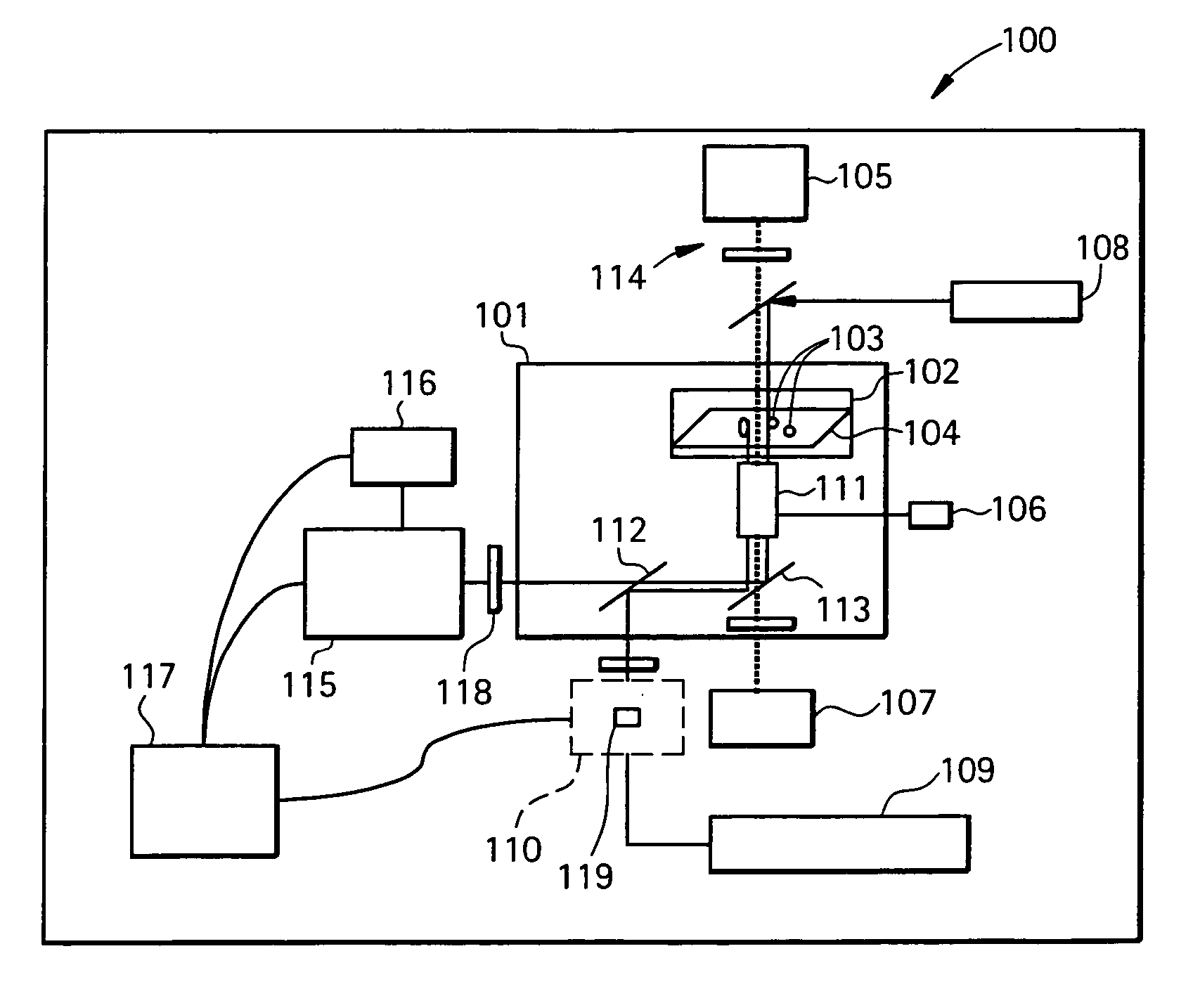
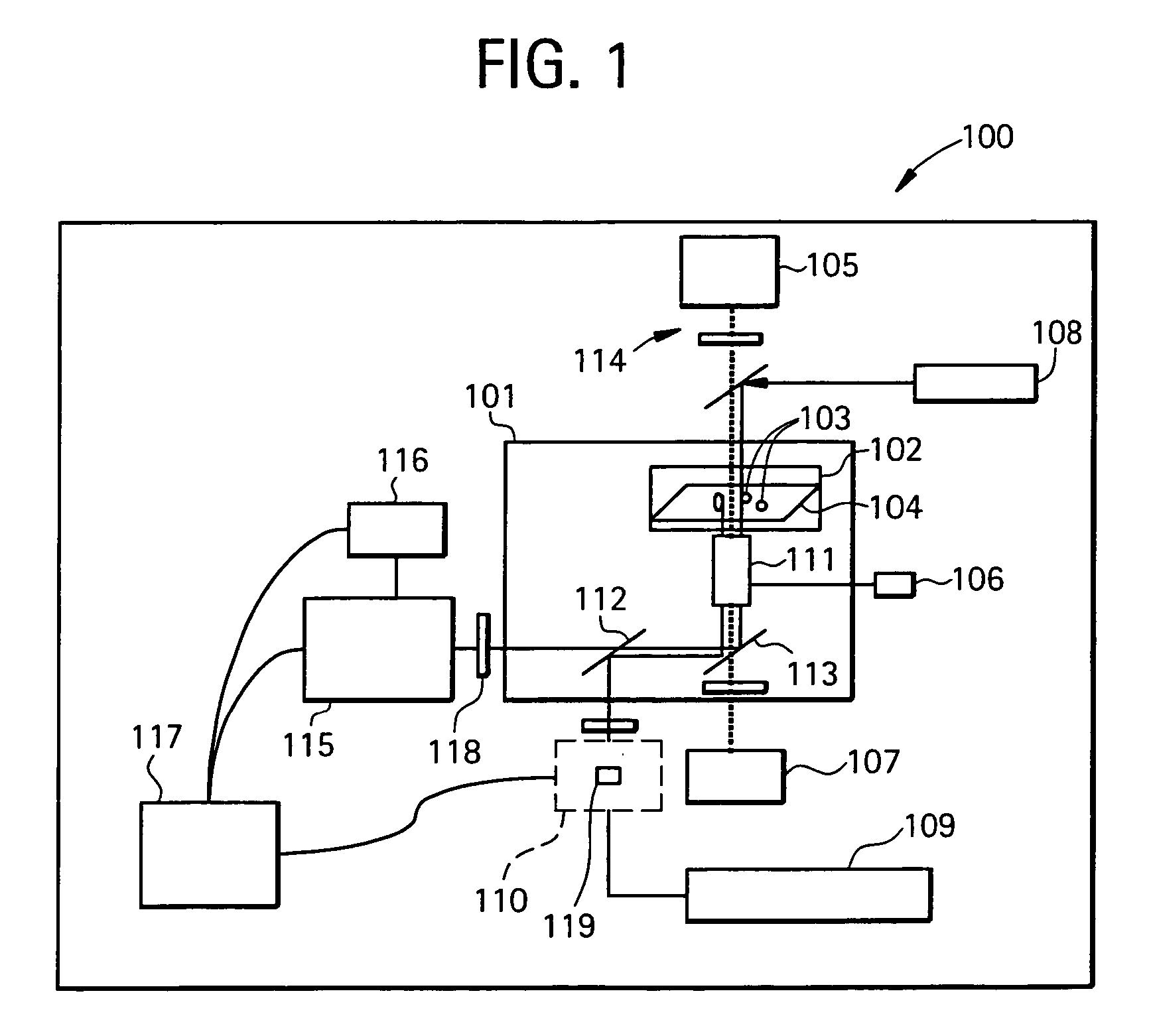
[0024] The present invention is related to an optical force measurement device that can apply optically based forces and sensitively detect the resulting deformations in objects, such as cells, while the objects rest on or adhere to a surface, rather than being in suspension as in previous applications.

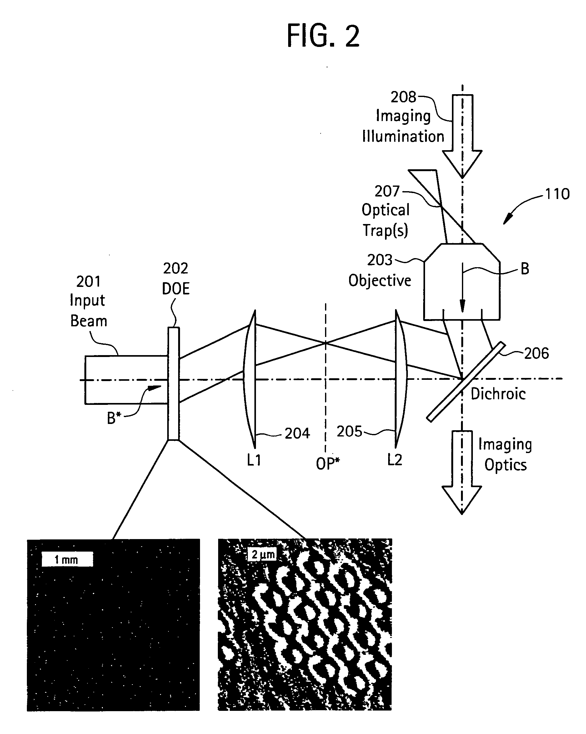
[0025] In particular, in one embodiment, the present invention measures deformations in the object as a function of an applied optical force, using holographic optical trapping technology, for example, to direct optical forces to potentially many regions of interest simultaneously.

[0026] In another embodiment, the present invention measures deformations in the object using a quantitative phase contrast microscopy technique—e.g. spatially modulated optical force microscopy (SMOFM).

[0027] In a preferred embodiment, the present invention utilizes spatially modulated optical force microscopy (SMOFM) with single beam optical force probing capability or with a holographic optical trappin...


PUM

PropertyMeasurementUnit
refractive index n2aaaaaaaaaa
center wavelengthaaaaaaaaaa

Abstract

The present invention utilizes spatially modulated optical force microscopy (SMOFM) with single beam optical force probing capability or with a holographic optical trapping system capable of multi-beam optical force probing coupled to a microscope objective, to generate a probe beam(s) as a force probe to perturb the object that is adhered or resting on a surface, so that deformations of the object may subsequently be quantified. This quantification is performed by imaging a sequence of four phase shifted replicas of the image using a computer-controlled spatial light modulator, and calculating the pixel by pixel optical path-length using existing algorithms. The change in optical path lengths, and consequently the viscoelastic or elastic response elicited, is an indication of damage or disease when the objects are cells. In another embodiment, the optical deformability of the cells may be measured and correlated with measurements of cytoskeletal / structural protein expression.

Description

[0001] The present invention claims priority from U.S. Provisional Patent Application No. 60 / 726,620, filed Oct. 17, 2005, the contents of which are herein incorporated by reference in its entirety.BACKGROUND OF THE INVENTION [0002] Diseases are often characterized by their unique molecular and / or genetic fingerprints. However, for many diseases, including cancer, this has yielded limited success; partly because there are many possible ways the molecular pathways in a cell may become pathological, there is much to learn. [0003] Cancer is still a leading killer in the United States, despite decades of focused research activity on the problem. However cells, aside from being biochemical and genetic entities, are also mechanical entities which have physical properties such as elasticity. Diseases which effect the cytoskeletal protein network of cells (i.e. the structural integrity of the cell), including cancer, should naturally yield cells with altered mechanical properties (e.g. elas...

Claims


Application Information

Patent Timeline
Patent Type & Authority Applications(United States)
IPC IPC(8): G01N31/00
CPCB82Y20/00B82Y35/00G01N21/6456G03H2001/0077G01Q30/04G01Q60/20G01Q20/02
Inventor AKCAKIR, OSMAN
Owner ARRYX INC
Why Eureka
Try Eureka