
Extraction device and medical suturing device set

a technology of suturing device and extraction device, which is applied in the field of extraction device and medical suturing device set, can solve the problems of difficult suture support by the snare loop, organ separation from the parietal, and limited use of experienced health care personnel

Inactive Publication Date: 2007-05-24
TYCO HEALTHCARE GRP LP
View PDF39 Cites 52 Cited by
  • Summary
  • Abstract
  • Description
  • Claims
  • Application Information

AI Technical Summary

Benefits of technology

[0005] The object of the present invention is to offer an extraction device and medical suturing device set that solves the aforementioned problems by readily handing over the suture, so that it can be handled even by inexperienced health care personnel.
[0007] In the present invention, the suture inserted into the organism is engaged by the engagement member of the extraction needle, so it can be drawn out of the organism at a place that is different from the insertion location. Because this makes it easy to hand over the suture, it can be handled even by inexperienced health care personnel.
[0008] In one invented extraction device, the extraction device is furnished with a mantle that forms a cavity that permits the extraction needle to pass entirely through so as to be exposed from the tip. In the present invention, it is possible to insert the extraction needle into the cavity of the mantle, thus making it possible to reduce the invasiveness to the body when drawing the extraction needle on which the suture is caught outside of the body.
[0010] In the present invention, it is possible to engage the suture that has been inserted into the organism by the engagement member of the extraction device, and draw it out of the organism, though the cavity of the extraction puncture needle, at a location that is different from the insertion location. This makes it easy to hand over the suture, so it can be handled even by inexperienced health care personnel.
[0012] In the present invention, it is possible to have a structure in which the hook elements are facing several different directions outwards, towards 2 outer directions, or towards 4 outer directions; there is no limitation of the peripheral positioning of hook elements, which thus makes it easy to engage the suture that has been disposed in an annular form.
[0014] In the present invention, provision is made for an extraction device, an insertion puncture needle, and a suture, which makes it easy to hand over the suture, so the desired organ and paries can be securely anchored with low invasiveness even by inexperienced health care personnel. It is also possible to draw out a single suture from multiple locations, thus, by taking this together and ligating it, it becomes possible to anchor the organ in a plane. Here, anchoring the organ in a plane means that, for example, the organ is anchored by suturing such that the suture forms a triangular or quadrilateral surface within the organ. When the organ is thus anchored, there is no separation within the plane of the organ from the paries.

Problems solved by technology

Generally, when a catheter is inserted transdermally into the stomach, gallbladder, or other organs, a problem may arise in that the organ separates from the paries such that the catheter cannot be inserted into the organ.
In a conventional medical suturing device set, two needles are used to hand over the suture, so when the relative positions of the needle tips within the organ after puncturing are not correct, or when there is insufficient space within the organ and the snare loop is deformed against the organ wall, it becomes difficult for the suture to be supported by the snare loop, so its use is limited to experienced health care personnel.

Method used


Image

  • Extraction device and medical suturing device set
  • Extraction device and medical suturing device set
  • Extraction device and medical suturing device set

Examples


embodiment 1

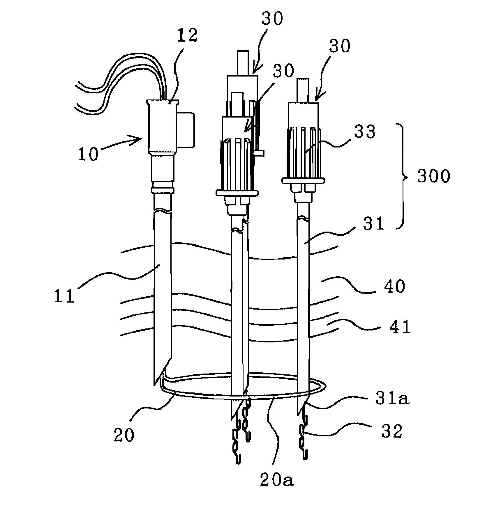
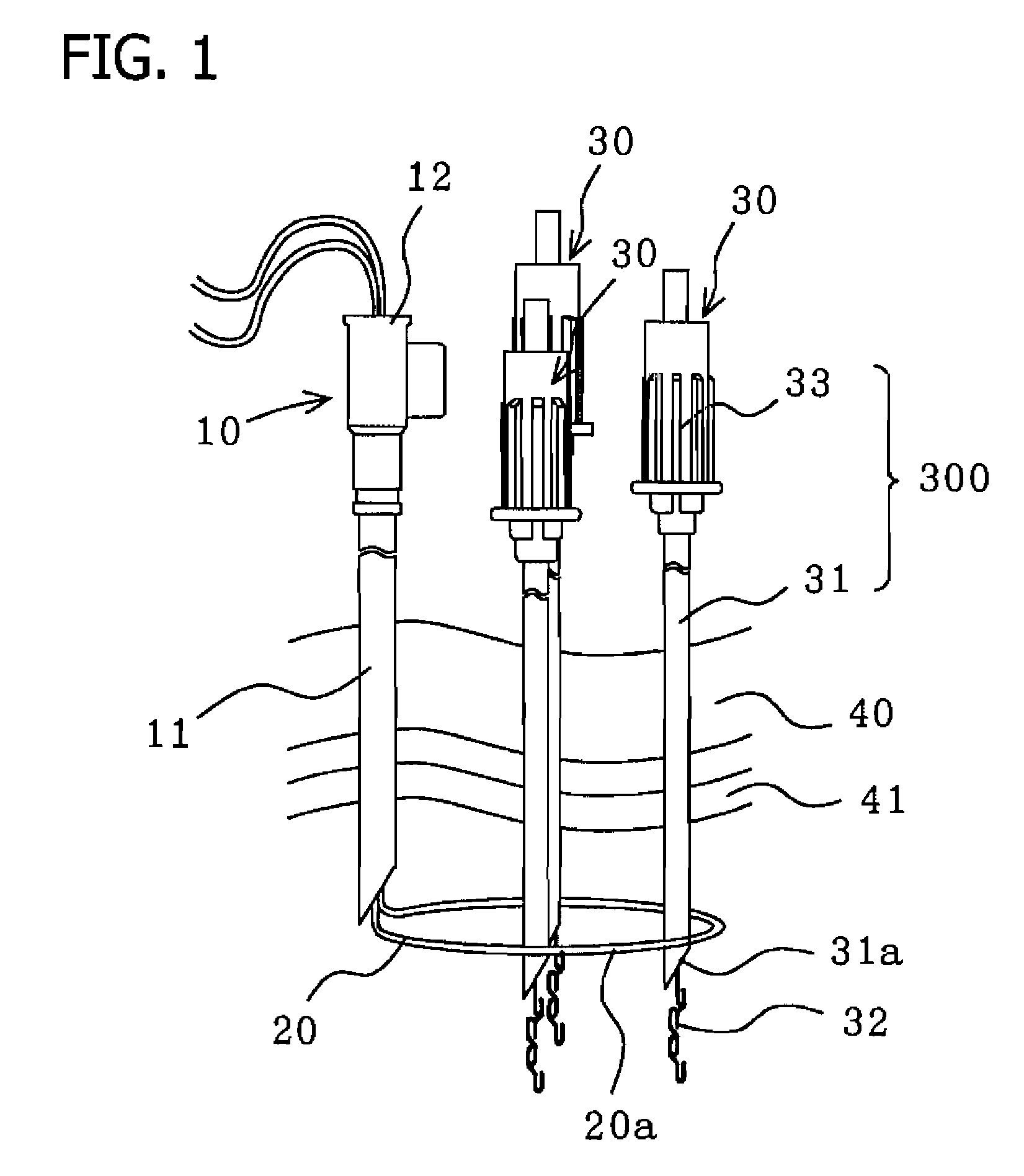
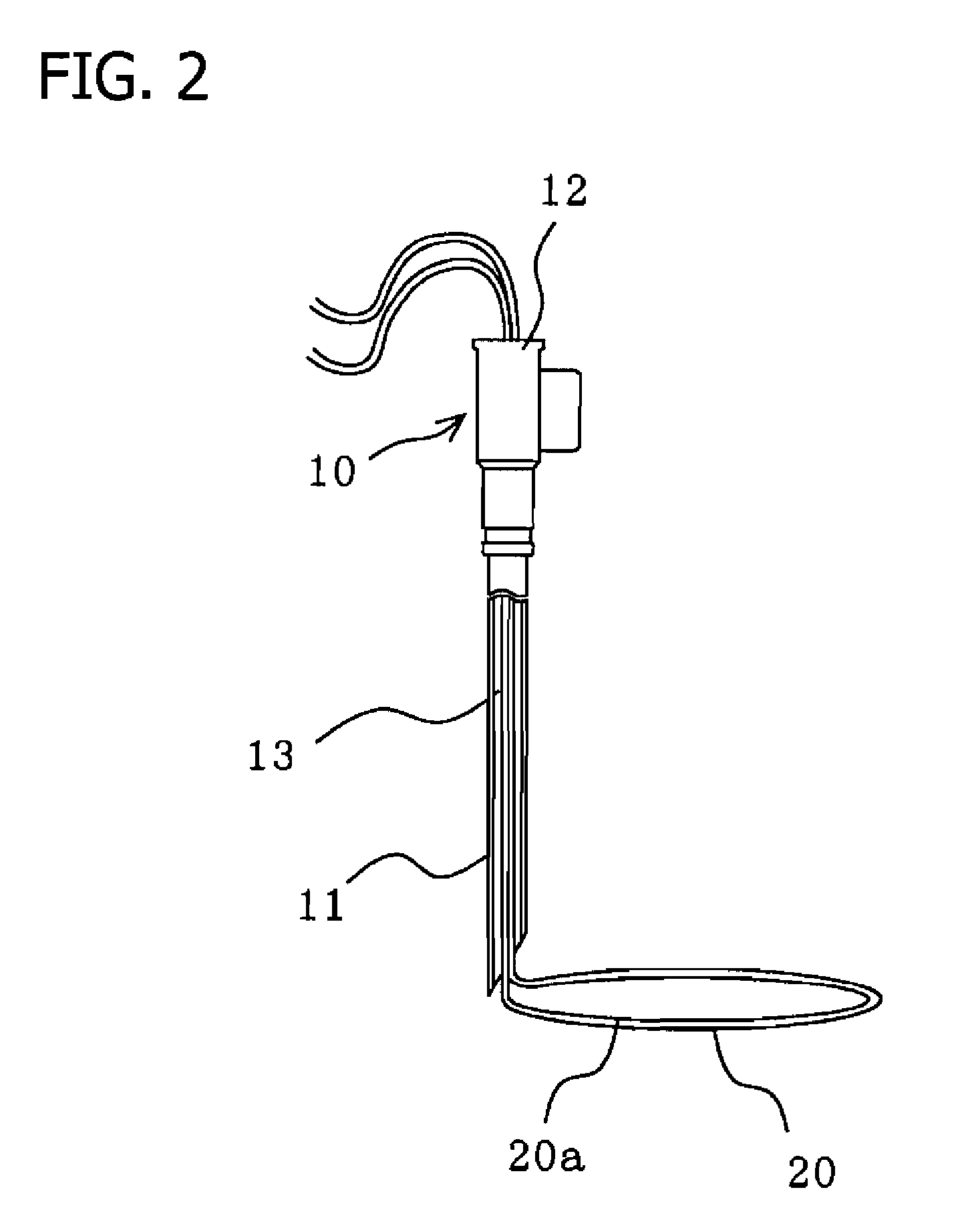
[0030]FIG. 1 is a side view of the medical suturing device set pertaining to Embodiment 1 of the present invention. FIG. 2 is a partially axially cut-away mode side view of the suture and insertion puncture needle of FIG. 1. FIG. 3 is a side view of the essential elements of the extraction device of FIG. 1. As shown in the figures, the medical suturing device set is structured by a insertion puncture needle 10 furnished with a suture 20, and several extraction devices 30 (3 in the illustrated example).

[0031] The insertion puncture needle 10 is structured using a puncture needle 11 and hub 12, with insertion through-hole 13 boring through these internally in the axial direction, such that suture 20 can slide within insertion through-hole 13. The puncture needle 11 is formed so that its apical tip has a sharp angle, and suture 20 protrudes from the apical tip so as to form a loop section 20a. The suture 20 may be constituted by a nylon thread, for example.

[0032] The extraction devic...

embodiment 2

[0042]FIG. 10 is a side view of the essential elements of the extraction device pertaining to Embodiment 2 of the present invention. In Embodiment 2, engagement member 52a of pulling out device 52 has a helical shape, so as to seize loop section 20a of suture 20 on helical engagement member 52a. More specifically, the engagement member 52a of pulling out device 52 is structured by a continuous helical element, and this helical section thrusts out from insertion puncture needle 10 so as to engage part of the formed loop section 20a.

[0043] According to Embodiment 2, the engagement member 52a of pulling out device 52 is formed as a helical shape, so without being limited to the circumferential position of pulling out device 52, it is easily able to engage the suture that is disposed in annular shape. Other than pulling out device 52, the structure, method of use, and results of Embodiment 2 are identical to those of Embodiment 1 so the explanation [of identical aspects] may be omitted...

embodiment 3

[0044]FIG. 11 is a side view of the essential elements of the extraction device pertaining to Embodiment 3 of the present invention. In Embodiment 3, engagement member 62a of pulling out device 62 has a paperclip shape, so that loop section 20a of suture 20 is seized on the paperclip-shaped section.

[0045] Here, pulling out device 62 is structured by a paperclip-shaped engagement section; for example, it may be formed by 3 U-shaped sections. More specifically, as shown in FIG. 11, a linear element has been bent on the side of metal needle 31 to form primary U-shaped element 620a; the vicinity of primary U-shaped element 620a has been bent on the side of primary U-shaped element 620a to form tertiary U-shaped element 622a; the vicinity of primary U-shaped element 620a has been further bent on the side of metal needle 31 to form secondary U-shaped element 621a; and the primary and secondary U-shaped elements 620a and 621a positioned in identical orientations with partial overlap and t...


PUM


Abstract

The extraction device is furnished with a pulling out device on which are formed hook elements that hook onto part of suture, which is disposed in an annular shape within the organism, and are able to draw this out of the organism. It is also a medical suturing device set that is used to anchor the organ to the organism's paries, which is furnished with a puncture needle, in which insertion through-hole is formed from the basal tip side to the apical tip side, and which is able to puncture the paries from the outside such that the aperture of insertion through-hole on the apical tip side is positioned within the organ; a suture that is supplied to the inside of the organ from the aperture via insertion through-hole, and which is able to from an annular section within the organ.

Description

[0001] The present invention pertains to an extraction device and medical suturing device set used when anchoring human organs and paries. [0002] Generally, when a catheter is inserted transdermally into the stomach, gallbladder, or other organs, a problem may arise in that the organ separates from the paries such that the catheter cannot be inserted into the organ. Therefore, it is necessary to anchor the organ and paries prior to catheter insertion and prevent the organ from separating from the paries. [0003] Recently, the mainstream of medical suturing device sets has become a device in which two needles are used to hand over the suture within an organ, and knot both ends of the suture drawn out of the organism, thus anchoring the organ and paries. Such a device comprises a suture, a suture insertion needle capable of inserting a suture into an organ, and an organ anchoring needle equipped with a tubular external needle and an internal needle having a tip equipped with a snare lo...

Claims


Application Information

Patent Timeline
IPC IPC(8): A61B17/04
CPCA61B17/0482A61B17/0483A61B17/0485A61B17/06066A61B17/06109A61B2017/00349A61B2017/0472A61B2017/06052A61B2017/061A61B2017/0619
Inventor FUNAMURA, SHIGEAKIMABUCHI, HITOSHI
Owner TYCO HEALTHCARE GRP LP
Learn More