
Cross-time and cross-modality inspection for medical image diagnosis

a cross-modality and imaging technology, applied in image analysis, medical/anatomical pattern recognition, instruments, etc., can solve the problems of inconsistent and correct interpretation of a series, difficult coordination of multiple images with respect to each other, and poor delineation of anatomy

Inactive Publication Date: 2007-10-11
CARESTREAM HEALTH INC
View PDF8 Cites 50 Cited by
  • Summary
  • Abstract
  • Description
  • Claims
  • Application Information

AI Technical Summary

Problems solved by technology

Positron emission tomography (PET) and single photon emission computed tomography (SPECT) scans display aspects of brain function and allow metabolic measurements but poorly delineate anatomy.
Unfortunately, such a coordination of multiple images with respect to each other is extremely difficult and even highly trained medical personnel, such as experienced radiologists, have difficulty in consistently and properly interpreting a series of medical images so that a treatment regime can be instituted which best fits the patient's current medical condition.
Another problem encountered by medical personnel is the large amount of data and numerous images that are obtained from current medical imaging devices.
In order for medical personnel to properly review each image takes a great deal of time and, with the many images that current medical technology provides, a great amount of time is required to thoroughly examine all the data.
However, Smith's technique fails to teach how to inspect cross-time, cross-modality medical images for a patient so that reliable and accurate diagnoses and surgical plan can be performed.
Daw's system does not provide a function to automatically detect and differentiate image areas corresponding to materials (tissues) being imaged that have different time response to contrast enhancing agent.
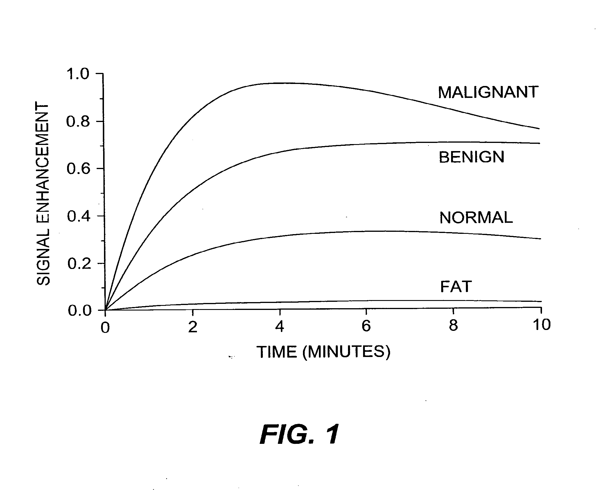
Studies of such curves / parameters has been used clinically to identify and characterize tumors into malignant or benign classes, although the success has been variable with generally good sensitivity but often very poor specificity (for example, refer to S. C. Rankin “MRI of the breast”, Br. J. Radiol 73, pp 806-818, 2000).

Method used

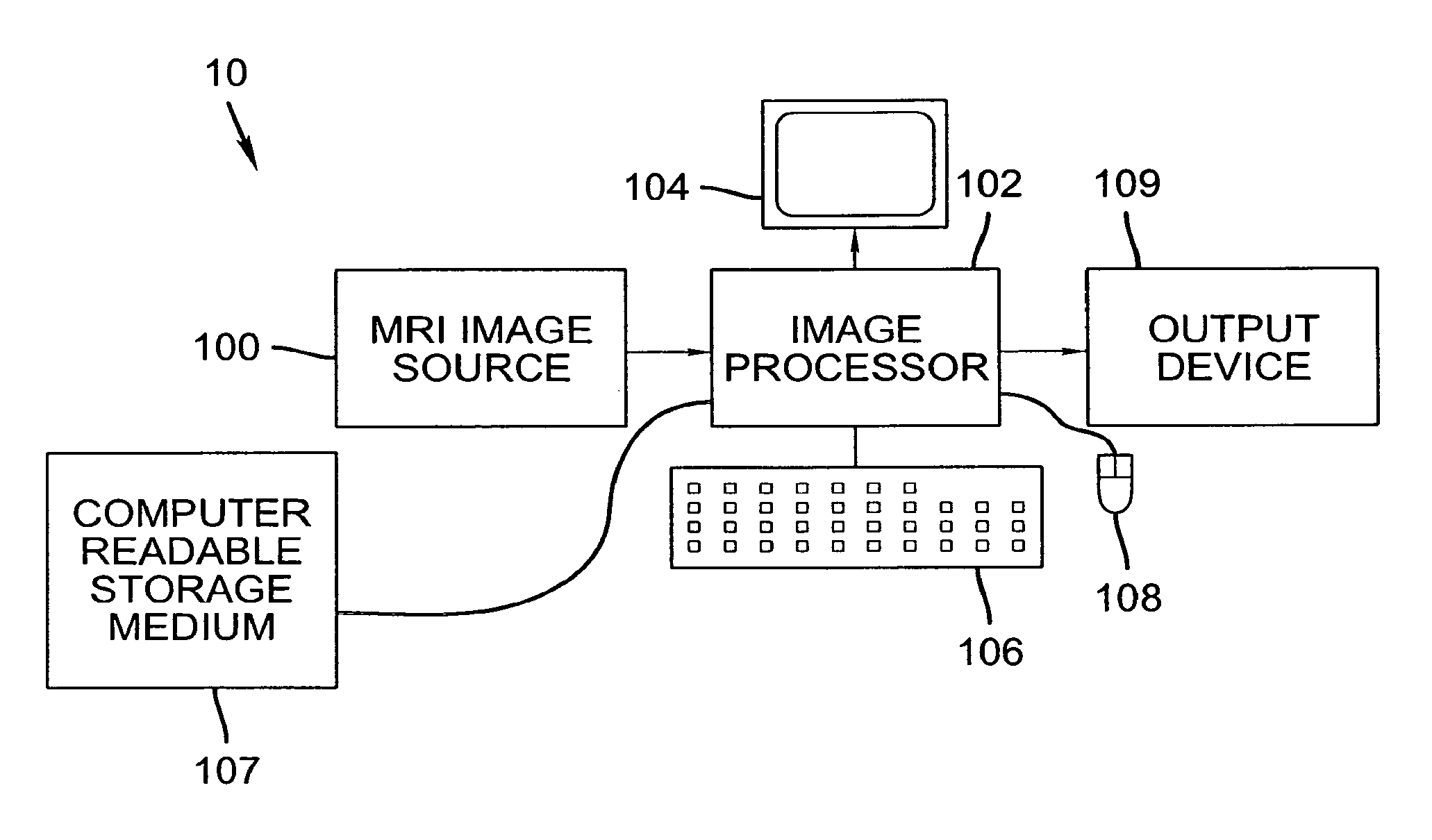
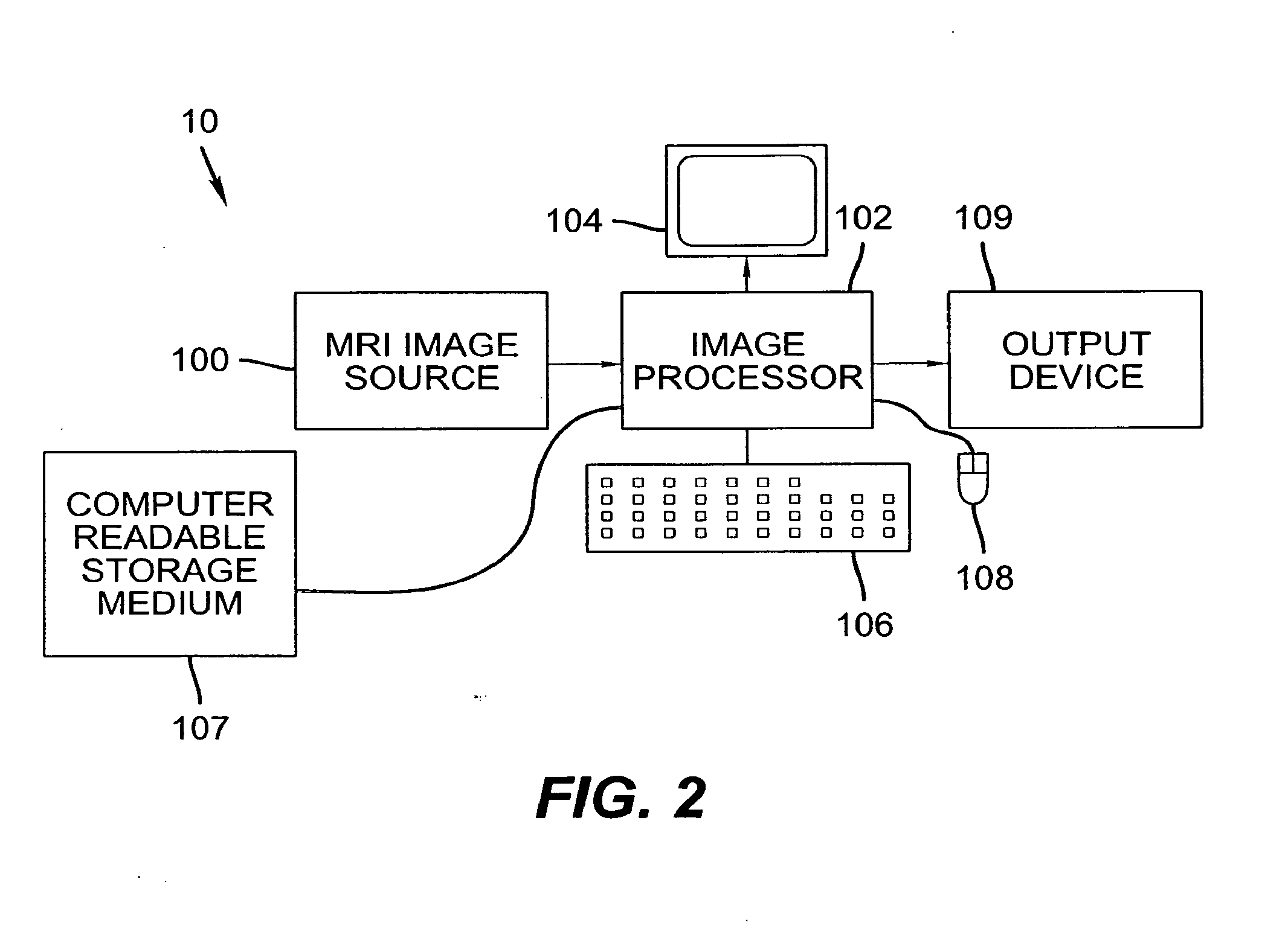

Image

  • Cross-time and cross-modality inspection for medical image diagnosis
  • Cross-time and cross-modality inspection for medical image diagnosis
  • Cross-time and cross-modality inspection for medical image diagnosis

Examples


Embodiment Construction

[0043] The following is a detailed description of the preferred embodiments of the invention, reference being made to the drawings in which the same reference numerals identify the same elements of structure in each of the several figures.

[0044] The inspection of cross-time, cross-modality medical images for a patient can assist in providing reliable and accurate diagnoses and surgical planning. For example, X-ray mammography has limited specificity and sensitivity. It is reported that 5%- 15% of cancers are missed using X-ray mammograms. MRI mammography, as an alternative imaging method, has a high sensitivity for tumors larger than 3 mm.

[0045] It is known that malignant breast tumors begin to grow their own blood supply network once they reach a certain size; this is the way the cancer can continue to grow. In a breast MRI scan, a contrast agent injected into the bloodstream can provide information about blood supply to the breast tissues; the agent “lights up” a tumor by highli...


PUM


Abstract

A cross-time and cross-modality inspection method for medical image diagnosis. A first set of medical images of a subject is accessed wherein the first set is captured at a first time period by a first modality. A second set of medical images of the subject is accessed, wherein the second set is captured at a second time period by a second modality. The first and second sets are each comprised of a plurality of medical image. Image registration is performed by mapping the plurality of medical images of the first and second sets to predetermined spatial coordinates. A cross-time image mapping is performed of the first and second sets. Means are provided for interactive cross-time medical image analysis.

Description

CROSS REFERENCE TO RELATED APPLICATIONS [0001] Reference is made to, and priority is claimed from, U.S. Provisional Patent Application No. 60 / 754,884, titled “CROSS-TIME AND CROSS-MODALITY INSPECTION FOR MEDICAL IMAGE DIAGNOSIS” in the names of Chen et al., provisionally filed on Dec. 29, 2005. [0002] Reference is made to U.S. Provisional Patent Application No. 60 / 755,156, titled “CROSS-TIME INSPECTION FOR MEDICAL IMAGE DIAGNOSIS” in the names of Chen et al., provisionally filed on Dec. 30, 2005.FIELD OF THE INVENTION [0003] The present invention relates to a digital image processing method for image analysis and, in particular, to cross-time and cross-modality inspection of tissues of different properties (for example, abnormal and normal tissues) in medical image. BACKGROUND OF THE INVENTION [0004] Digital imaging techniques for medical applications were implemented in the 1970′s, for example, with the clinical use of the Computed Tomography (CT) scanner. Since then, use of x-ray ...

Claims


Application Information

Patent Timeline
IPC IPC(8): G06K9/00
CPCG06K9/6203G06T7/0038G06K2209/05G06K9/6269G06T7/38G06V10/7515G06V2201/03G06F18/2411
Inventor CHEN, SHOUPUHUO, ZHIMINRAY, LAWRENCE A.
Owner CARESTREAM HEALTH INC
Learn More