Medical device introduction systems and methods

a technology for medical devices and introduction systems, applied in the field of medical device introduction systems and methods, can solve the problems of limiting the ability of physicians to perform as well as capable, limiting the ability of physicians to view a target site, limiting the ability of construction of medical devices, and limiting the ability of physicians to perform additional procedures. to achieve the effect of improving steering

Inactive Publication Date: 2008-03-06
OPTIVIA MEDICAL
View PDF17 Cites 256 Cited by
  • Summary
  • Abstract
  • Description
  • Claims
  • Application Information

AI Technical Summary

Benefits of technology

[0011] In some embodiments, one or more of the introducer tube, the working channel tube, and the endoscopic cannula can include a proximal portion having a first durometer and a distal portion having a second durometer. The second durometer is lower t

Problems solved by technology

As a result, operating within small spaces with a plurality of medical devices, such as scopes, dilating and cutting instruments, fluids, catheters, implants, and the like, can become difficult to manage.
When performing a procedure with a plurality of medical devices, positioning, controlling, manipulating, and handling the various medical devices during the procedure can limit a physician's ability to perform as well as capable.
That is, the design and construction of a medical device can limit a physician's ability to view a target site, maneuver within a space, transition between procedures, and/or perform additional procedures.
Managing the use of multiple devices in a procedure can pose even greater difficulty to a single physician who desires to perform a procedure, often without assistance or with limited assistance, in an office or outpatient setting so as to avoid the time and expense of hospital utilization for such procedures.
The optical capabilities can be limited due to various factors, including, for example, the anatomical structures about which the scopes are maneuvered, and the movement and/or control together of both the imaging device and a delivery device and the resulting loss in orientation in an interior body region.
For example, optical capabilities with conventional endoscopes typically used in hysteroscopy procedures are often limited in such ways, making it difficult for the physi

Method used


Image

  • Medical device introduction systems and methods
  • Medical device introduction systems and methods
  • Medical device introduction systems and methods

Examples


Embodiment Construction

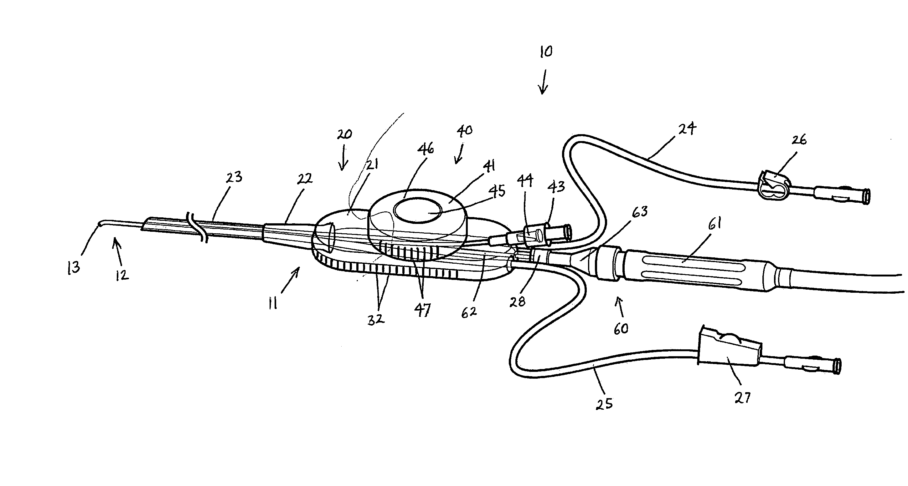
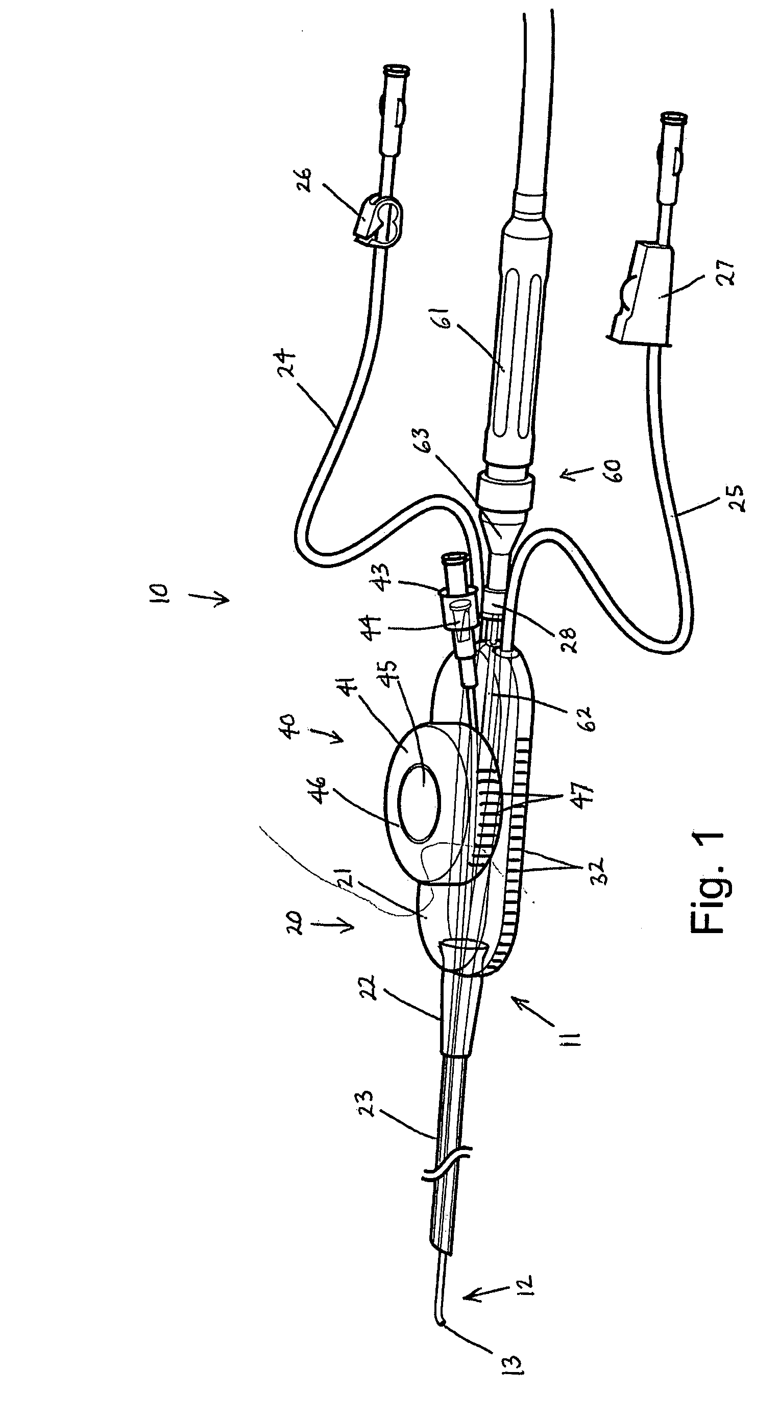
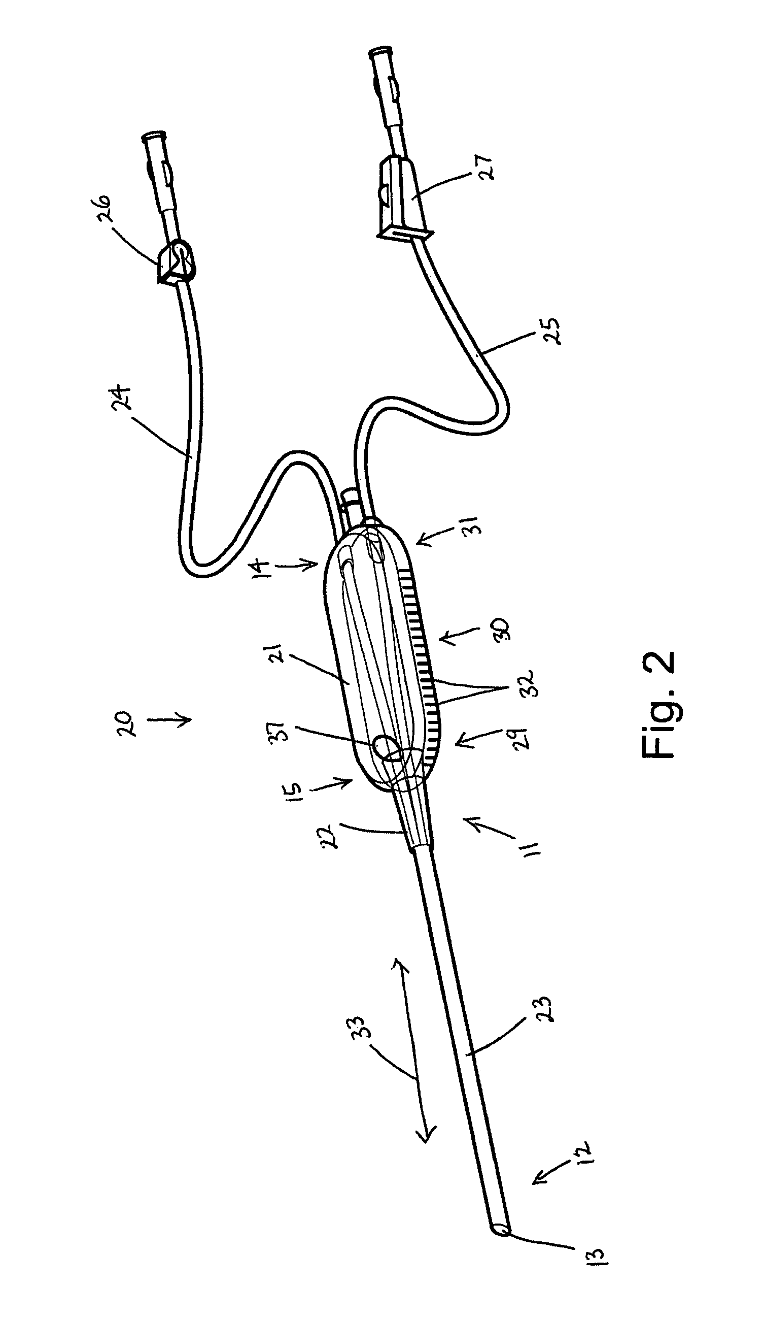
[0037] Some embodiments of the present invention can provide a medical device introduction system and / or method. FIGS. 1-18 show various aspects of such embodiments. For example, an illustrative embodiment of a medical device introduction system and / or method can include a medical introducer, a separate imaging device, and / or a separate working channel device. In such an embodiment, each of the medical introducer, the imaging device, and the working channel device can be movable independent of the other.

[0038] Minimally invasive surgical procedures have been developed that can be used in many diagnostic and / or therapeutic medical procedures. Such minimally invasive procedures can reduce pain, post-operative recovery time, and the destruction of healthy tissue. In minimally invasive surgery, the site of pathology can be accessed through portals rather than through a significant incision, thus preserving the integrity of intervening tissues. These minimally invasive techniques also o...


PUM


Abstract

A medical device introduction system and method can include a medical introducer, a separate imaging device, and/or a separate working channel device, each of which may be movable independent of the other. The medical introducer can include a handle and an elongate introducer tube extending from the handle and having a plurality of lumens, and may be inserted into an interior body region of a patient. The separate imaging device may be inserted through the handle and positioned in one of lumens. The separate working channel device can include an elongate working channel tube and a position controller. The working channel tube can include at least one lumen defining a working channel. The position controller can be configured to control positioning of the working channel tube. The working channel device may be removably connected to the handle and positioned in another lumen.

Description

CROSS-REFERENCE TO RELATED APPLICATIONS [0001] This application claims benefit of U.S. Provisional Patent App. No. 60 / 813,807, filed Jun. 14, 2006, and U.S. Provisional Patent App. No. 60 / 878,230 filed Jan. 3, 2007, each of which is incorporated by reference herein in its entirety.FIELD OF THE INVENTION [0002] The present invention relates to medical device introduction systems and methods. Embodiments of the present invention may be useful for separately introducing and independently controlling multiple cooperating medical devices in interior body regions. BACKGROUND OF THE INVENTION [0003] In recent years, medical procedures have advanced to stages where less and less invasive, or minimally invasive, surgeries, diagnostic procedures, exploratory procedures, or other medical procedures have been desired and demanded by patients, physicians, and payers. To accomplish these desires and demands, various medical devices and instrumentation have been developed, such as cannulas or micr...

Claims


Application Information

Patent Timeline
IPC IPC(8): A61B1/00
CPCA61B1/00135A61B1/0051A61B1/018A61B2017/003A61B1/303A61B17/42A61B1/07A61B1/01
Inventor SNOKE, PHILLIP JACKMASSEY, JOE B.FINCH, MARCUS E.MCMILLAN, JOHN A.MARTEL, MARK CURRANCASSIDY, KENNETH TODDALLRED, PHILIPWILSON, JON S.ROTHSTEIN, LAWRENCE B.
Owner OPTIVIA MEDICAL
Why Eureka
Try Eureka