
Cardiac Valve Data Measuring Method And Device

a technology of cardiac valves and data measuring devices, applied in the field of cardiac valve data measuring methods and devices, can solve the problems of difficult three-dimensional analysis and measurement through use of three-dimensional images, deterioration of cardiac function, and difficult to find the anatomical and positional relations between the mitral valves

Inactive Publication Date: 2008-04-10
YD +4
View PDF1 Cites 36 Cited by
  • Summary
  • Abstract
  • Description
  • Claims
  • Application Information

AI Technical Summary

Benefits of technology

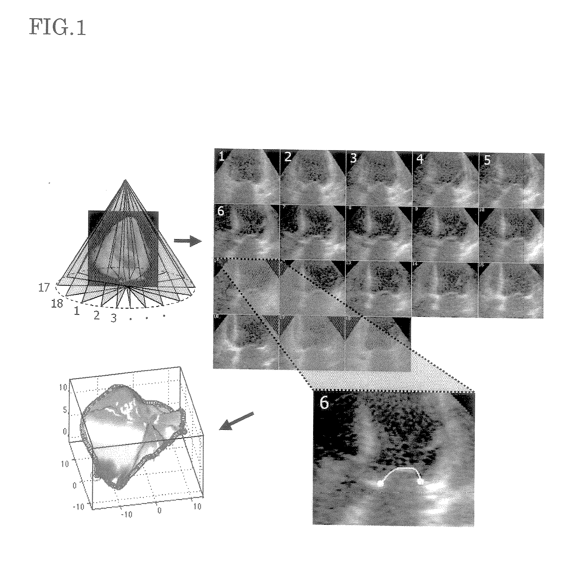
[0027] The device of the present invention has the following advantageous features (1) The device can provide three-dimensional display and quantitative analysis of the mitral valve complex, which have been impossible when conventional two-dimensional echocardiograms are employed. (2) Whereas the conventional reconstruction of a three-dimensional image from two-dimensional images is a laborious and time-consuming process, the device of the present invention requires only a short time (currently, about 15 minutes) before completion of the entire process, including three-dimensional analysis of the mitral valve (collection of echo images, tracing of the images, reconstruction of a three-dimensional image, and quantitative analysis of three-dimensional data).
[0028] Three-dimensional quantitative analysis using a three-dimensional echocardiogram has never been realized, and the present inventors are the first in the world to have accomplished the present invention. In particular, at present, attention is drawn worldwide to the elucidation of the mechanism of “functional mitral regurgitation” which is caused by the malfunction of the papillary muscle or the left ventricle even though the cusp and leaflet of the mitral valve have no anomaly, as well as to the development of therapy therefor. Studies on these themes, which have relied on analysis of two-dimensional echocardiogram images, are expected to greatly progress because three-dimensional analysis has become possible. The device of the present invention can be used for preoperative diagnosis and surgical treatment of mitral regurgitation, and is therefore clinically very useful.

Problems solved by technology

However, such an operation causes various problems after replacement with an artificial valve, such as deterioration of the cardiac function, and complication associated with an anticoagulation treatment.
However, in the echocardiography widely used at the present, diagnosis is performed by use of a two-dimensional image, and therefore, it has been difficult to find the anatomical and positional relations between the mitral valve, which has a complex three-dimensional structure, and the surroundings thereof.
However, three-dimensional analysis and measurement through use of a three-dimensional image is still difficult, and the actual specific configuration and positional relation cannot be quantized.
Therefore, presently, three-dimensional echocardiography has not been put in actual use for clinical purposes.

Method used


Image

  • Cardiac Valve Data Measuring Method And Device
  • Cardiac Valve Data Measuring Method And Device
  • Cardiac Valve Data Measuring Method And Device

Examples


Embodiment Construction

[0037] A best mode for carrying out the present invention will be described with reference to the drawings In the description hereinbelow, the following abbreviations and acronyms will be used. [0038] MR=mitral regurgitation [0039] 3D=three-dimensional [0040] 2D=two-dimensional [0041] LV=left ventricle [0042] LA=left atrium [0043] ROA=regurgitation orifice area [0044] EF=ejection fraction [0045] PISA=proximal isovelocity surface area [0046] EDV=end-diastolic volume (capacity) [0047] ESV=end-systolic volume (capacity)

[0048] Two-dimensional echocardiography provides the following examinations. When standard 2D echocardiography is performed for all subjects, the end-diastolic volume (EDV) and end-systolic volume (ESV) of each subject can be measured by a modified Simpson's method (wherein the entire left ventricle is approximated as a stack of cylinders). As a result, the ejection fraction (%) can be calculated by an equation 100×(EDV−ESV) / EDV. MR is evaluated by color Doppler echocar...


PUM


Abstract

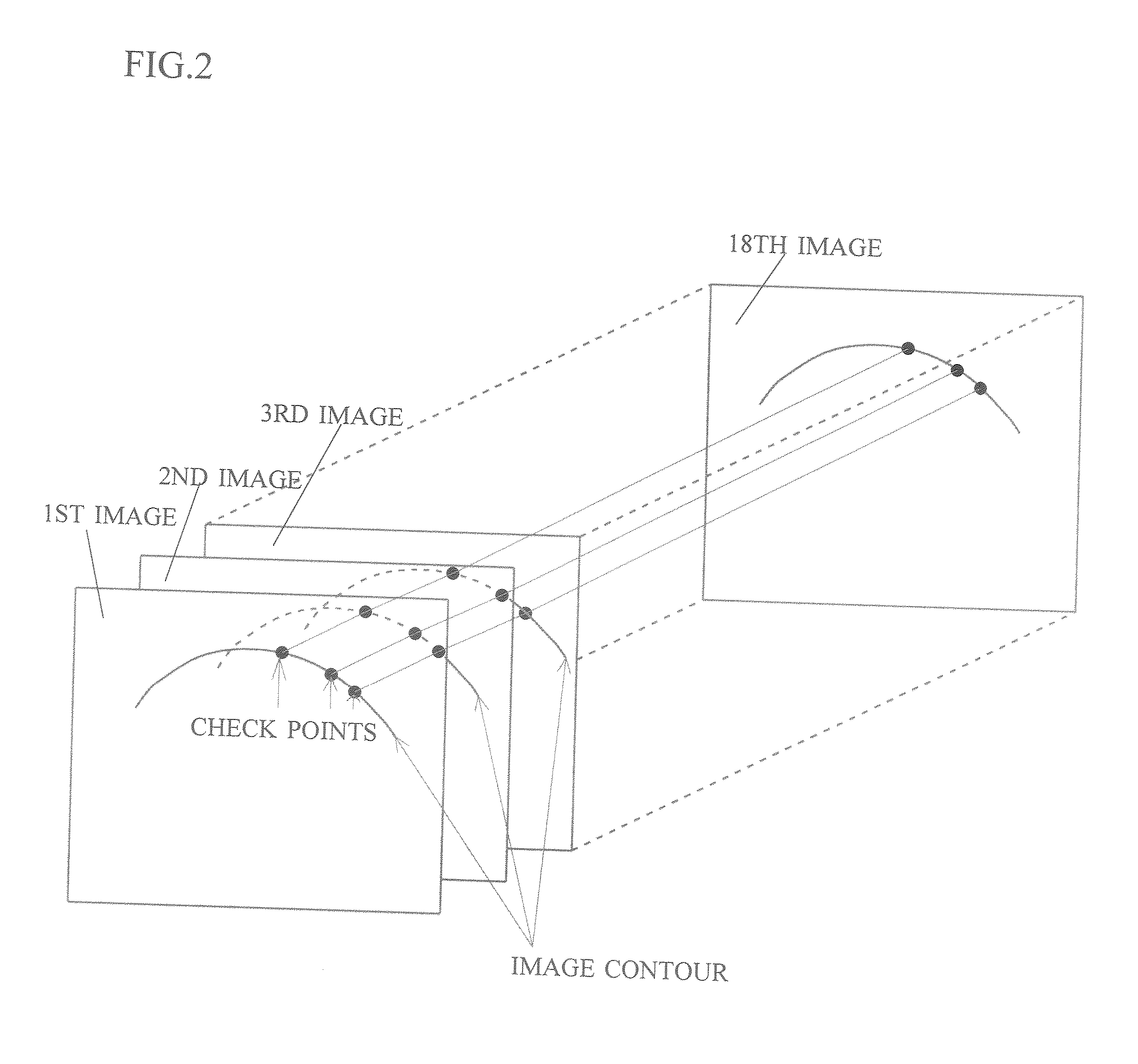
An object of the present invention is to acquire information regarding the cardiac valve required in the clinical field, such as the tenting volume, tenting area, tenting height of the mitral valve of the heart, the area, the circumferential length, and height (the difference between the highest portion and the lowest portion) of the mitral annulus, etc. A method of obtaining a three-dimensional cardiac-valve image for measuring clinically required data regarding the cardiac valve, in which method a three-dimensional echocardiogram is created from two-dimensional echocardiograms obtained through scanning by means of an echocardiograph, and the three-dimensional cardiac-valve image is automatically extracted from the three-dimensional echocardiogram by computer processing The method is characterized in that a fitting evaluation function (potential energy) of a model of the mitral annulus in a fitting model prepared in consideration of the physical shapes of the heart and the mitral annulus is optimized by the replica exchange method and extended simulated annealing method.

Description

TECHNICAL FIELD [0001] The present invention relates to a method and device for obtaining measurements or data of a cardiac valve to be used for clinical purposes More specifically, the present invention relates to a method and device for automatically extracting a clear three-dimensional image of a cardiac valve, from which various measurements or data regarding the cardiac valve can be obtained. BACKGROUND ART [0002] Mitral regurgitation (mitral valve insufficiency) frequently occurs among valvular diseases and in the case of severe regurgitation, left-sided cardiac failure occurs. Therapy for a severe mitral regurgitation is basically a surgical treatment and conventionally, a mitral-valve replacement operation using an artificial valve has been performed. However, such an operation causes various problems after replacement with an artificial valve, such as deterioration of the cardiac function, and complication associated with an anticoagulation treatment. Therefore, in recent y...

Claims


Application Information

Patent Timeline
Patent Type & Authority Applications(United States)
IPC IPC(8): G06K9/46
CPCA61B5/1075A61B8/08A61B8/0883A61B8/13G06T2207/30048G06T7/0083G06T7/0089G06T2207/10136G06T2207/20116A61B8/483G06T7/12G06T7/149A61B8/00
Inventor WATANABE, NOZOMIOGASAWARA, YASUOSAKURAI, MASASHI
Owner YD
Learn More