Modeling Cerebral Aneurysms in Medical Images

a technology of cerebral aneurysms and medical images, applied in image enhancement, angiography, instruments, etc., can solve the problems of adversely affecting the accuracy of the results to be used in treatment planning, and parameter setting is also not user-friendly

Active Publication Date: 2008-10-09
SIEMENS HEALTHCARE GMBH
View PDF19 Cites 35 Cited by
  • Summary
  • Abstract
  • Description
  • Claims
  • Application Information

AI Technical Summary

Problems solved by technology

Unfortunately, these techniques are often sensitive to parameter settings, and may adversely affect...

Method used


Image

  • Modeling Cerebral Aneurysms in Medical Images
  • Modeling Cerebral Aneurysms in Medical Images
  • Modeling Cerebral Aneurysms in Medical Images

Examples


Embodiment Construction

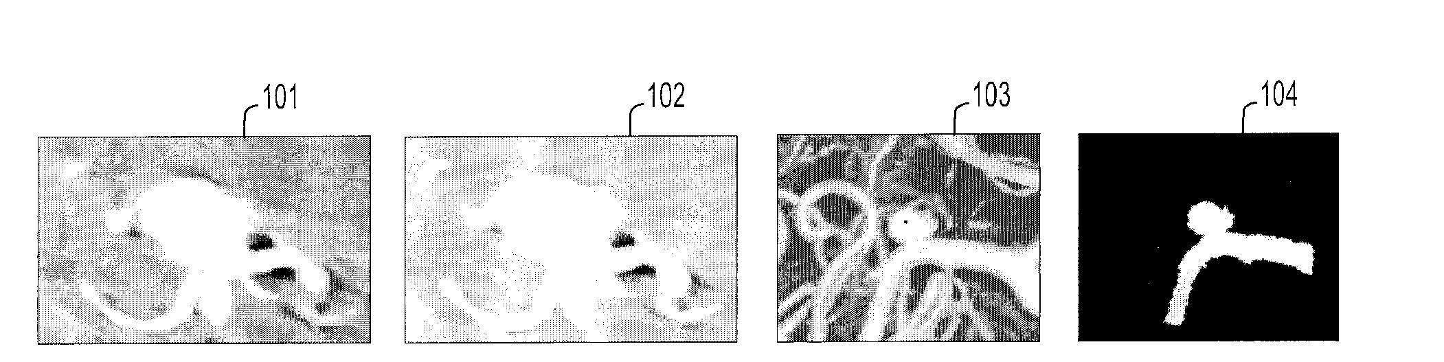
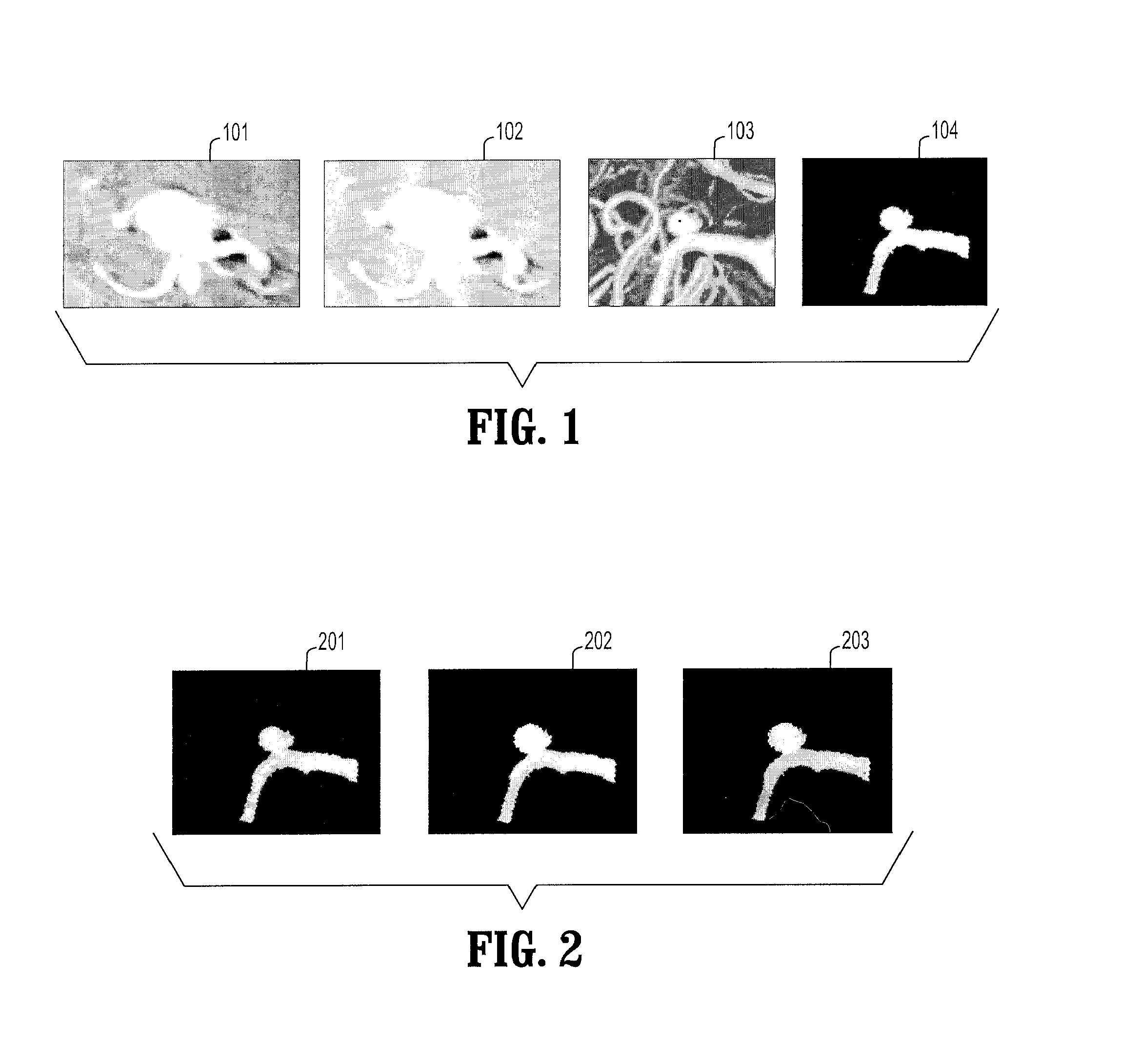
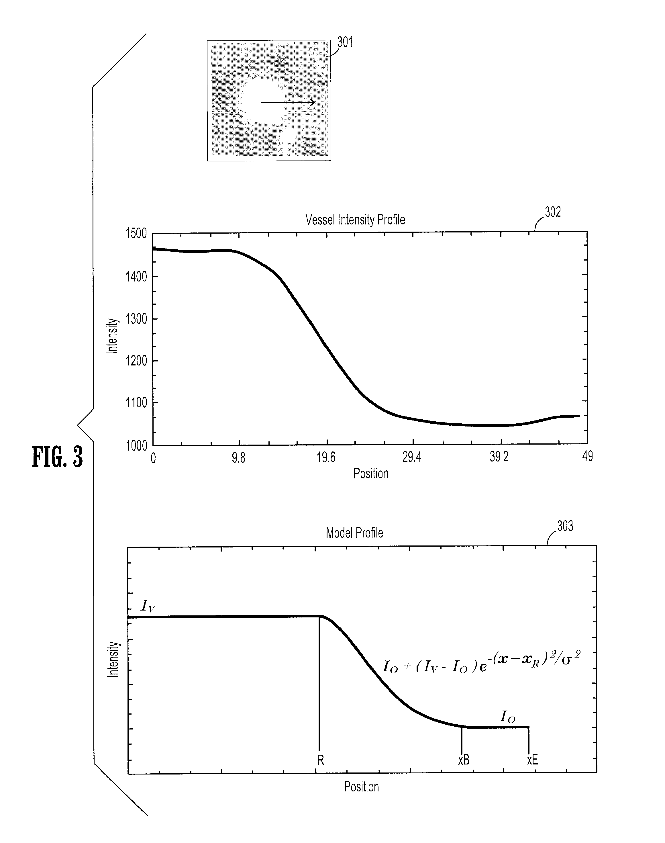
[0029]A new method for modeling aneurysms and their vessels, for instance, from 3D X-ray, MRA and subtraction CTA data via a single seed placement inside an aneurysm will be provided. A new method based on the principles of edge based graph cuts will be provided for the segmentation of aneurysms and their parental vessels, which delineates the boundary between vascular structures (aneurysms and vessels) and background accurately. However, when vascular structures are close to each other, they cannot be well separated due to the presence of strong partial voluming effects. As an aspect of the present invention, vessels will be accurately modeled up to aneurysms. A new framework for the extraction of center-axis representation as well as explicit vessel surface models from for instance 3D-X Ray, CTA / MRA data is provided. In this framework, local vessel-axis models are extracted by an algorithm which uses cross-sectional boundary models in a graph based optimization. The surface models...


PUM


Abstract

Methods and systems for modeling cerebral aneurysm and their incoming and outgoing vessels from 3D image data are disclosed. Aneurysms and vessels are segmented from their background using a graph-cuts method. Begin and end of vessels are determined. Construction of a centerline of the incoming and outgoing vessels using a measure of vesselness in calculating a minimum cost path in a graph with nodes being representation of pixels is also disclosed. Vessel surface models are constructed from sub-voxel cross-sectional segmentation. The interpolation of vessels inside an aneurysm based on smooth continuity is disclosed. Selection of endo-vascular stents based on interpolation results is also provided.

Description

CROSS-REFERENCE TO RELATED APPLICATIONS[0001]This application claims the benefit of U.S. Provisional Application No. 60 / 909,724 filed Apr. 3, 2007, which is incorporated herein by reference.BACKGROUND OF THE INVENTION[0002]The present invention relates to segmentation and visualization of blood vessels in image data. In particular it relates to modeling of cerebral aneurysms and incoming and outgoing vessels from 3D image data.[0003]Cerebral aneurysms are the enlargements of arteries, which often tend to occur at or near bifurcations of arteries of the brain. In recent years, non-invasive treatment of cerebral aneurysms has become popular due to the advances in image acquisition devices. In clinical settings, threshold based segmentation algorithms or maximum intensity projection (MIP) visualizations combined with user interactions are heavily used for the treatment planning of cerebral aneurysms.[0004]Unfortunately, these techniques are often sensitive to parameter settings, and ma...

Claims


Application Information

Patent Timeline
IPC IPC(8): G06G7/60
CPCA61B5/02014G06T7/0012G06T7/0081G06T7/0091G06T7/0093G06T7/60G06T2207/10072G06T2207/20044G06T2207/30016G06T2207/30101G06T2207/30172G06T7/11G06T7/155G06T7/162
Inventor TEK, HUSEYINGULSUN, MEHMET AKIF
Owner SIEMENS HEALTHCARE GMBH
Why Eureka
Try Eureka