System and Method for Quasi-Real-Time Ventricular Measurements From M-Mode EchoCardiogram

a ventricular measurement and quasi-real-time technology, applied in the field of quasi-real-time ventricular measurement of ventricular dimensions from mmode echocardiograms, can solve the problems of obstructing the real-time requirement, not meeting the accuracy required for clinical practice, and taking more than 2 months to perform all these image warping operations, so as to achieve fast and accurate derivation of ventricular measurements

Active Publication Date: 2008-11-13
SIEMENS MEDICAL SOLUTIONS USA INC
View PDF31 Cites 11 Cited by
  • Summary
  • Abstract
  • Description
  • Claims
  • Application Information

AI Technical Summary

Benefits of technology

[0017]Exemplary embodiments of the invention as described herein generally include methods and systems for a fast and accurate derivation of ventricular measurements from M-mode echocardiogram that combines local landmark detectors with global template detectors in a probabilistic, hierarchical, and discriminative manner. A learning approach for measuring ventricular dimensions from M-mode echocardiograms in quasi-real time localizes a cohort of landmarks, leading to a high-dimensional searching task. To efficiently explore the high-dimensional parameter space, local landmark detectors and global anatomic structure detectors are fused in a probabilistic, hierarchical, and discriminative manner. Experiments on a set of 89 images record a median absolute error of 0.57 mm in localizing the landmarks while taking about 300 ms to process each image on a standard personal computer with a 3 GHz XEON CPU and 3 GB of memory.

Problems solved by technology

Empirical evidence shows a certain effectiveness of the local approach, but it does not meet the accuracy required for clinical practices.
This has a potential of yielding accurate results but renders a search in a high-dimensional parameter space, which obstructs the real time requirement.
Performing all these image warping operations can take more than 2 months.
As described earlier, the local approach is fast but inaccurate while the global approach is accurate but slow.
One idea is to fuse the local and global approaches, however the challenge is how to combine them in a nontrivial manner such that both speed and accuracy can be guaranteed.

Method used


Image

  • System and Method for Quasi-Real-Time Ventricular Measurements From M-Mode EchoCardiogram
  • System and Method for Quasi-Real-Time Ventricular Measurements From M-Mode EchoCardiogram
  • System and Method for Quasi-Real-Time Ventricular Measurements From M-Mode EchoCardiogram

Examples


Embodiment Construction

[0037]Exemplary embodiments of the invention as described herein generally include systems and methods for quasi-real time measurements of ventricular dimensions from M-mode echocardiograms. Accordingly, while the invention is susceptible to various modifications and alternative forms, specific embodiments thereof are shown by way of example in the drawings and will herein be described in detail. It should be understood, however, that there is no intent to limit the invention to the particular forms disclosed, but on the contrary, the invention is to cover all modifications, equivalents, and alternatives falling within the spirit and scope of the invention.

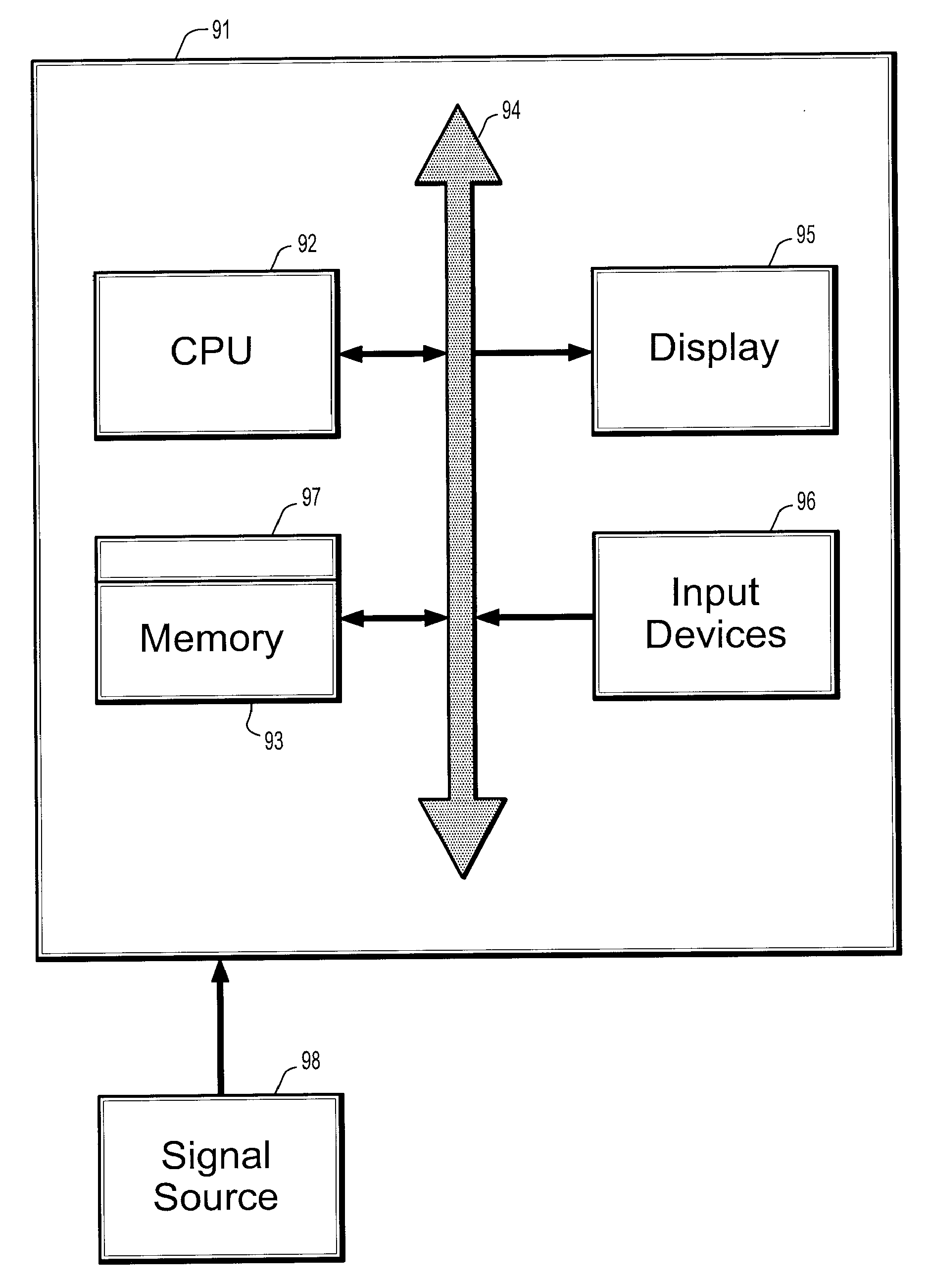
[0038]As used herein, the term “image” refers to multi-dimensional data composed of discrete image elements (e.g., pixels for 2-D images and voxels for 3-D images). The image may be, for example, a medical image of a subject collected by computer tomography, magnetic resonance imaging, ultrasound, or any other medical imaging syst...


PUM


Abstract

A method for measuring ventricular dimensions from M-mode echocardiograms, includes providing a digitized M-mode echocardiogram image, running a plurality of local classifiers, where each local classifier trained to detect a landmark on either an end-diastole (ED) line or an end-systole (ES) line in the image, recording all possible landmarks detected by the classifiers, where a search range in an N-dimensional parameter space defined by the landmarks for each dimension is reduced to a union of subsets, where each dimension of the parameter space corresponds a landmark, for each combination of possible landmarks, checking if an order of the landmarks is consistent with a known ordering of the landmarks, and if the order is consistent, running a global detector on each consistent combination of landmarks to find a landmark combination with a highest detection probability as a confirmed landmark detection, where the landmarks are used for measuring ventricular dimensions.

Description

CROSS REFERENCE TO RELATED UNITED STATES APPLICATIONS[0001]This application claims priority from “Accurate and Quasi-real-time Ventricular Measurements from M-mode Echocardiogram”, U.S. Provisional Application No. 60 / 908,208 of Zhou, et al., filed Mar. 27, 2007, the contents of which are herein incorporated by reference in their entirety.TECHNICAL FIELD[0002]This disclosure is directed to quasi-real time measurements of ventricular dimensions from M-mode echocardiograms.DISCUSSION OF THE RELATED ART[0003]M-mode echocardiogram is a spatial-temporal image slice of the human heart captured by an ultrasound device. Unlike regular B-mode echo that uses multiple interrogation beams, the M-mode echo uses a single interrogation beam and hence achieves an enhanced temporal and spatial resolution along a single line. It is often used in clinical practice to assess the functionality of anatomic structures inside the heart, such as the left ventricle and aortic root, as its high image quality a...

Claims


Application Information

Patent Timeline
Patent Type & Authority Applications(United States)
IPC IPC(8): A61B8/00
CPCA61B5/1075A61B5/7267A61B6/503A61B8/08A61B8/0883A61B8/463G06K9/00523G06K9/00536G06K9/6257G06T7/60A61B8/486G16H50/70G06V2201/031G06V10/7747G06F18/2148G06F2218/08G06F2218/12
Inventor ZHOU, SHAOHUA KEVINGUO, FENGJACKSON, JOHNBRENDEL, MICHAELCOMANICIU, DORIN
Owner SIEMENS MEDICAL SOLUTIONS USA INC
Why Eureka
Try Eureka