Pericardial space imaging for cardiac support device implantation

a technology for pericardial space and cardiac support, which is applied in the direction of prosthesis, application, ultrasonic/sonic/infrasonic diagnostics, etc. it can solve the problems of limited access approaches that cannot effectively show the location of other structures such as the a-v groove, the coronary sinus, the atrial appendage, etc., and the location of these csd structures with respect to those of the heart can be difficult to assess

Active Publication Date: 2008-12-25
DIAXAMED LLC
View PDF20 Cites 10 Cited by
  • Summary
  • Abstract
  • Description
  • Claims
  • Application Information

AI Technical Summary

Problems solved by technology

Unfortunately, limited access approaches may not permit direct intraoperative visualization of the CSD.
Fluoroscopic visualization can demonstrate the general shape of the heart and the location of the heart apex, but may not effectively show the location of other structures such as the A-V groove, the coronary sinus, the atrial appendages or the location of the pulmonary artery or right ventricular outflow tract.
Although structures of the CSD may be radioopaque, and visible through fluoroscopic imaging, the locations of these CSD structures with respect to those of the heart can be difficult to assess.
Accurate placement of the CSD can therefore be difficult to assess.

Method used


Image

  • Pericardial space imaging for cardiac support device implantation
  • Pericardial space imaging for cardiac support device implantation
  • Pericardial space imaging for cardiac support device implantation

Examples


Embodiment Construction

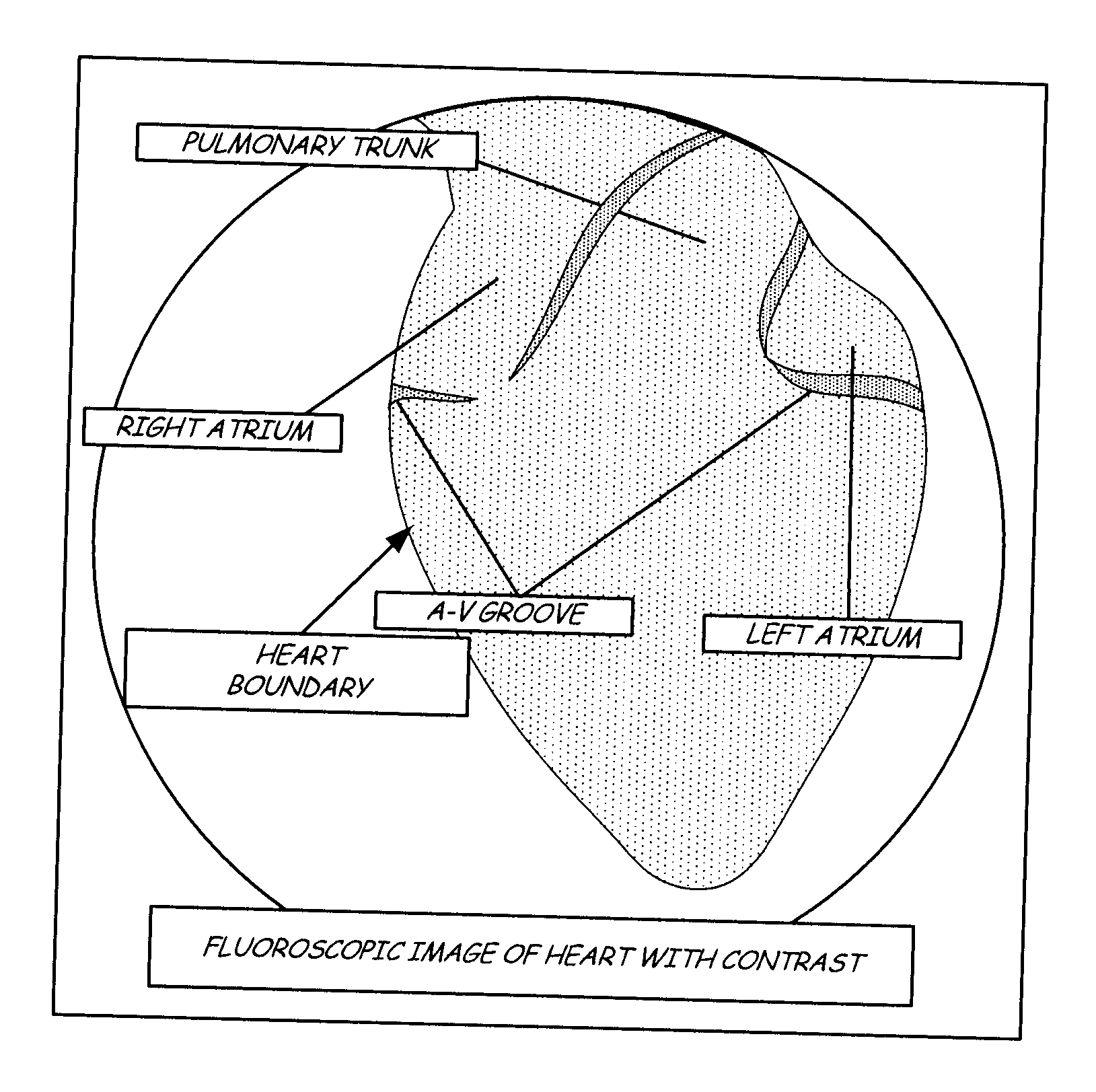
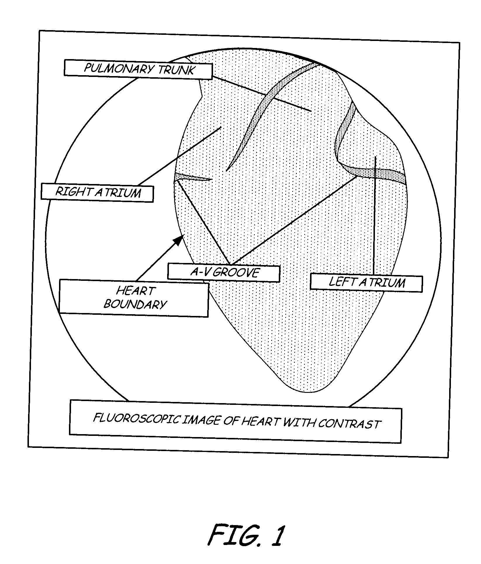
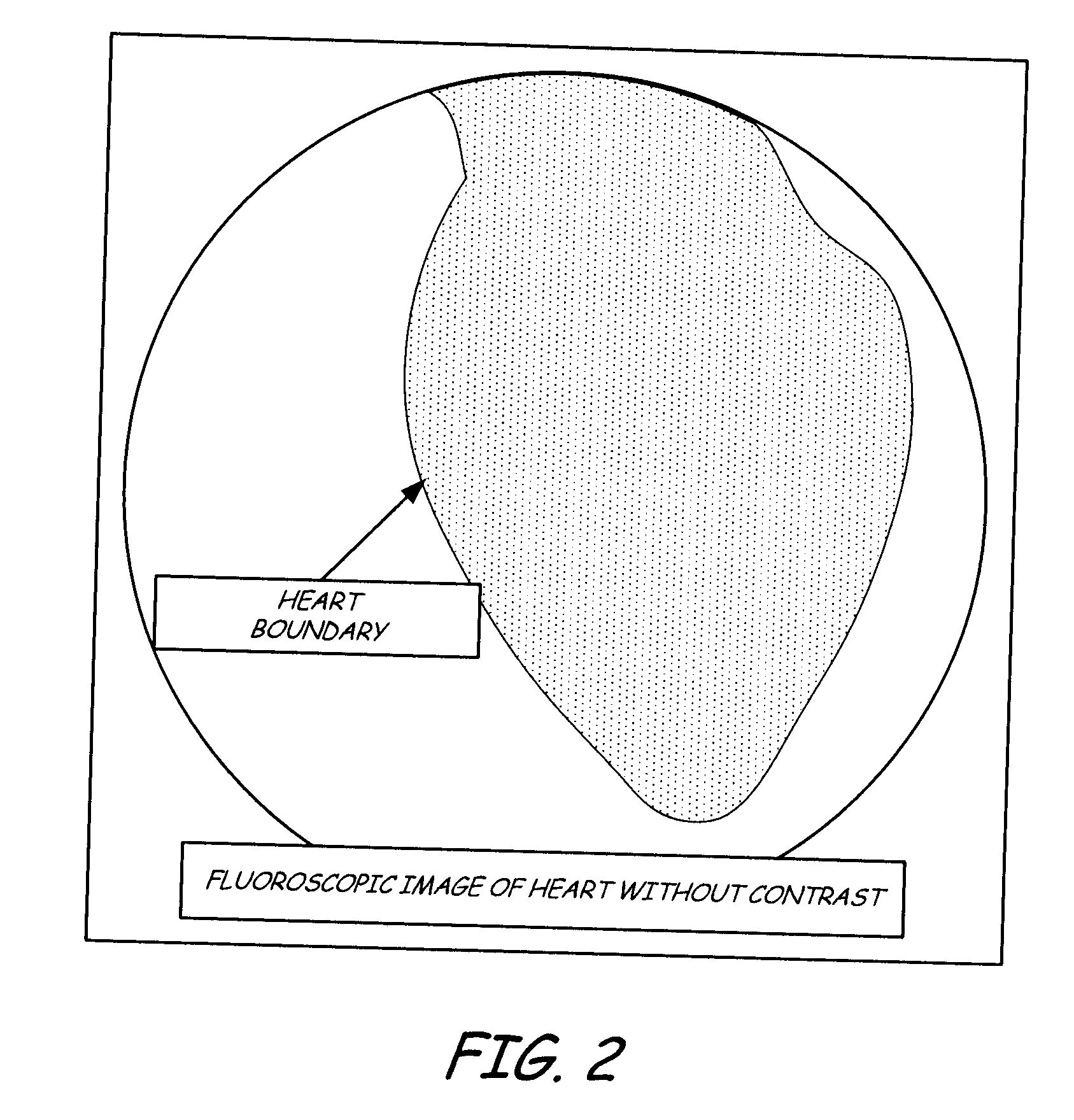
[0011]When an amount of iodinated or other contrast agent solution (e.g., up to about 30 cc in one embodiment) is introduced into the intact pericardial space, the vigorous motion of the heart distributes the contrast rapidly throughout the pericardial space. The contrast collects in groves and fissures on the surface of the heart. This includes the crevice between the left atrial and right atrial appendages and adjacent structures, as well as the space between the aorta and superior vena cava, and the crevice adjacent to the coronary sinus. These contrast-filled crevices are located adjacent to the coronary sulcus and the atrioventricular (A-V) groove, and provide radiographic markers that effectively demonstrate where these structures on the heart are located. A CSD can then be placed accurately on the heart. Irrigating and evacuating the pericardial sack repeatedly with normal saline can remove the contrast at the end of a procedure, if so desired. Alternatively, the contrast can...


PUM


Abstract

A method for implanting a cardiac support device (CSD) on a patient's heart. An amount of contrast agent sufficient to cause structures on the heart to be visible upon fluoroscopic or other imaging is introduced into the pericardial space surrounding the heart. The heart and contrast agent are imaged to provide a visual indication of the location of the structures of the heart. The CSD is placed on the heart using the visual indications provided by the imaging.

Description

REFERENCE TO RELATED APPLICATION[0001]This application claims the benefit of U.S. Provisional Application Ser. No. 60 / 936,594 filed on Jun. 21, 2007, and entitled “Pericardial Space Imaging For Cardiac Support Device Implantation,” which is incorporated herein by reference in its entirety.FIELD OF THE INVENTION[0002]The invention relates to methods for implanting cardiac support devices.BACKGROUND OF THE INVENTION[0003]Cardiac support devices are structures, sometimes referred to as jackets, that surround all or portions of a diseased heart. These devices are intended to treat chronic heart failure or other cardiac disease, which may be associated valvular dysfunction, by constraining expansion of the heart. They can be delivered and implanted using conventional cardiothoracic surgical techniques or minimally invasive surgical procedures. Devices of these types and associated delivery tools and methods are shown, for example, in the following U.S. patents and published applications,...

Claims


Application Information

Patent Timeline
Patent Type & Authority Applications(United States)
IPC IPC(8): A61F2/00
CPCA61B6/12A61B6/481A61M5/007A61B6/503A61B8/0883A61B6/487
Inventor COHN, WILLIAM E.HJELLE, AARON J.WALSH, ROBERT G.BASSETT, KEVIN PAUL
Owner DIAXAMED LLC
Why Eureka
Try Eureka