Imaging endoscope

a technology of endoscopes and endoscopes, which is applied in the field of imaging endoscopes, can solve the problems of high cost, low resolution, and inability to design single-use or disposable endoscopes

Inactive Publication Date: 2009-01-08
SCI MED LIFE SYST
View PDF23 Cites 88 Cited by
  • Summary
  • Abstract
  • Description
  • Claims
  • Application Information

AI Technical Summary

Problems solved by technology

One problem with conventional imaging endoscopes is their relatively low resolution.
Another problem is their high cost.
At a current price of approximately $350 each, the cost of such image sensors alone makes it impractical to design single use or disposable endoscopes.
However, the same factors that contribute to the long life of an endoscope also reduce its ability to be inserted into some body cavities.

Method used


Image

  • Imaging endoscope
  • Imaging endoscope
  • Imaging endoscope

Examples


Embodiment Construction

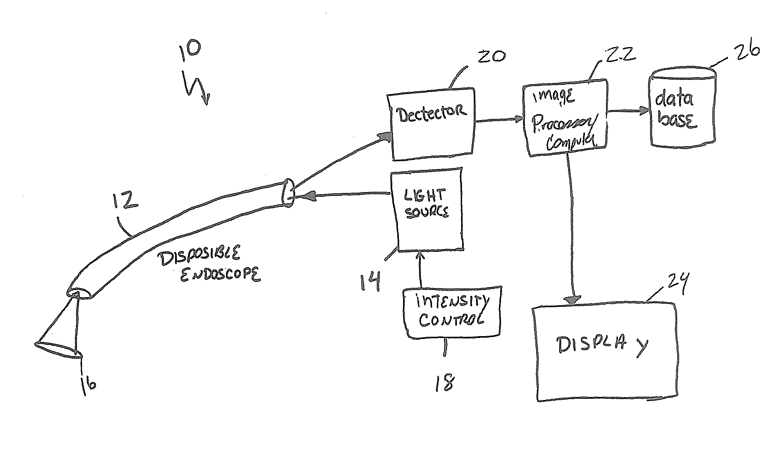
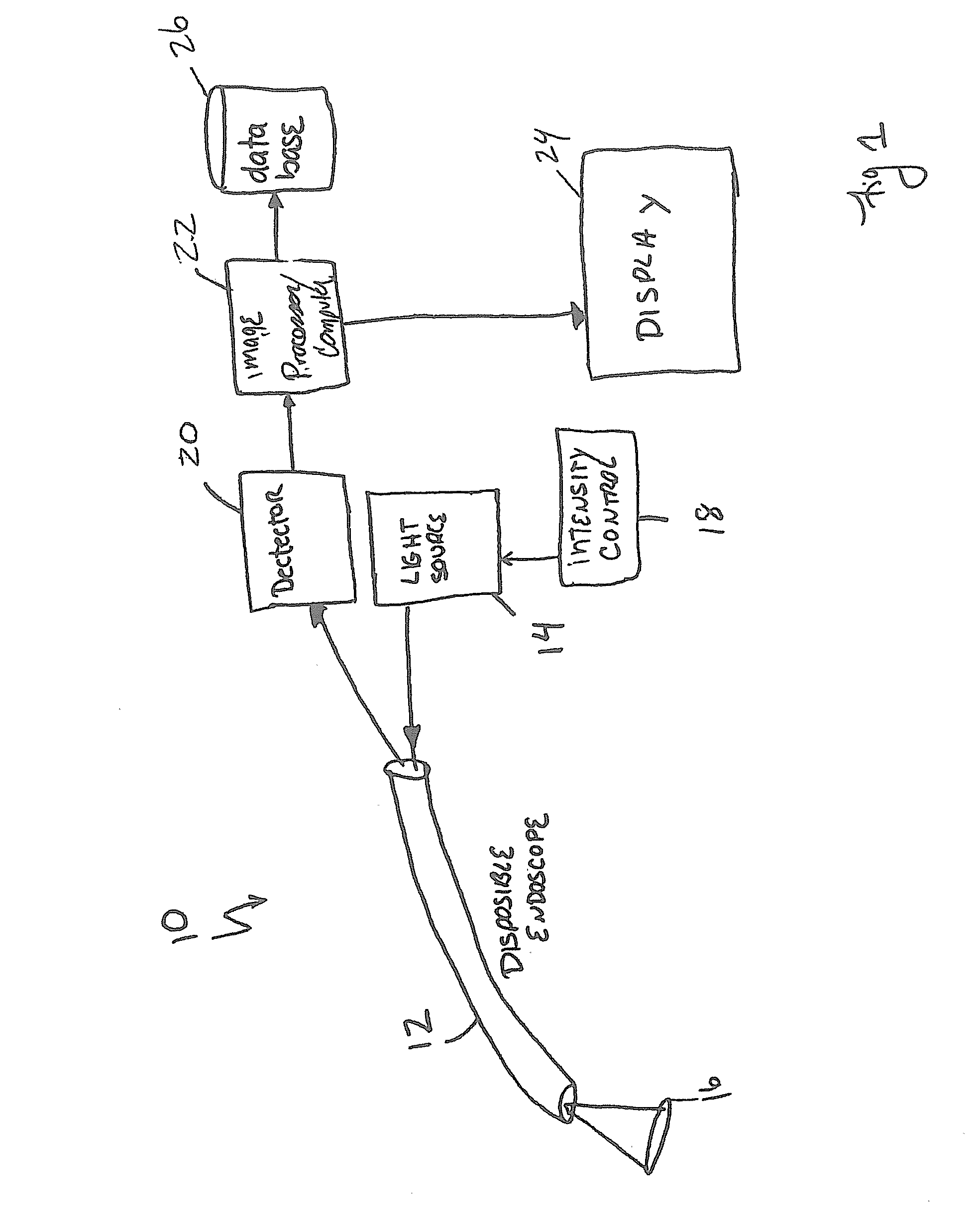
[0011]FIG. 1 illustrates one embodiment of a disposable imaging system 10 in accordance with the present invention. The imaging system 10 includes a disposable endoscope 12 generally comprising an elongate tube that directs light from a light source 14 onto an area of interest 16 that is within an internal body cavity (not shown). Light reflected from the area of interest 16 is gathered and returned through the endoscope 12 to a photo detector 20. The photo detector 20 generates electronic signals that are proportional to the intensity of the received light. The electronic signals produced by the photo detector 20 are supplied to an image processor / computer 22 that combines the electronic signals produced over the area of interest and creates an image of the tissue. Images produced by the image processor / computer 22 are displayed on a display device 24 such that a physician or other user can view the internal body tissue of a patient. The images from the image processor may be recor...


PUM


Abstract

An endoscopic imaging system includes an endoscope with a beam deflecting mechanism at or adjacent its distal end for directing a beam of illumination light over an area of interest. Reflected light is gathered by one or more lenses and supplied to a light sensor and an image processor / computer that produces an image of the tissue. In one embodiment, the beam deflecting mechanism comprises a pair of mirrors that are oscillated such that light is scanned in a raster pattern over the area of interest.

Description

FIELD OF THE INVENTION[0001]The present invention relates to medical devices in general, and in particular to imaging endoscopes.BACKGROUND OF THE INVENTION[0002]One of the most common methods for non-invasively screening an internal body cavity of a patient is with an imaging endoscope. Such endoscopes are elongated devices that are inserted into the body cavity. Light is delivered through an illumination channel of the endoscope and reflected light is gathered by one or more lenses that are coupled to an imaging channel. Light from the imaging channel is transmitted out of the endoscope and supplied to a camera or other viewing device so that a physician can examine the internal body tissue. Typical cameras connected to the endoscope typically include a solid state image sensor such as a CCD array.[0003]One problem with conventional imaging endoscopes is their relatively low resolution. For example, on a 3.5 mm square CCD array, the resolution is limited to approximately 850K pixe...

Claims


Application Information

Patent Timeline
Patent Type & Authority Applications(United States)
IPC IPC(8): A61B1/07A61B5/00
CPCA61B1/00103A61B1/07A61B1/00096A61B5/0084A61B5/0062A61B1/0627
Inventor BANIK, MICHAEL S.
Owner SCI MED LIFE SYST
Why Eureka
Try Eureka