System and Method for Organ Segmentation Using Surface Patch Classification in 2D and 3D Images

Active Publication Date: 2009-04-09
SIEMENS HEATHCARE GMBH
View PDF10 Cites 28 Cited by
  • Summary
  • Abstract
  • Description
  • Claims
  • Application Information

AI Technical Summary

Benefits of technology

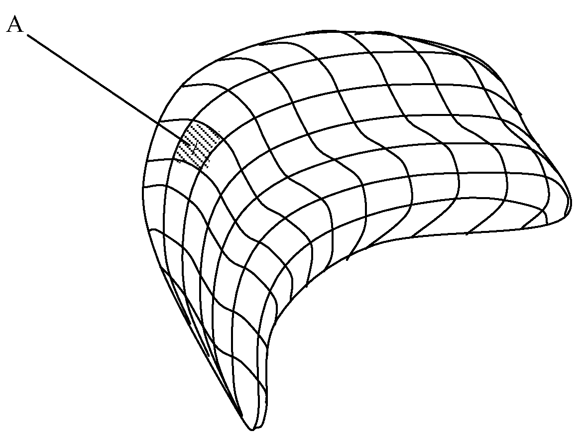
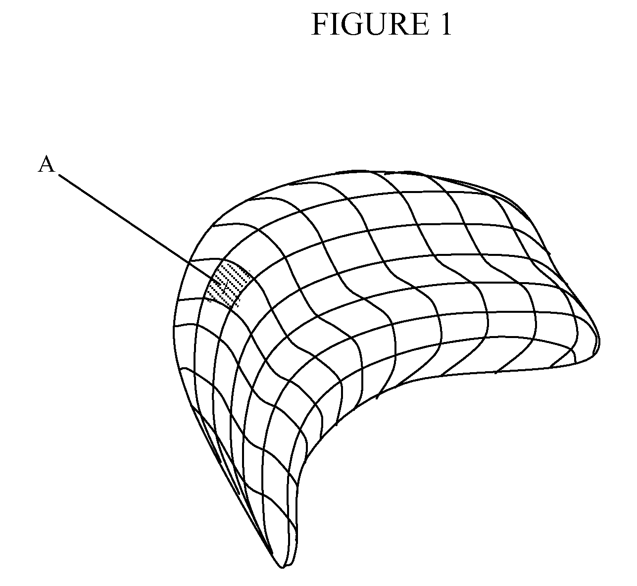
[0020]According to a further aspect of the invention, the positive and negative image feature examples are calculated in a direction orthogonal to the surface mesh by calculating, for each mesh cell aro

Problems solved by technology

The 3-dimensional datasets obtained from scanning modalities such as computed tomography (CT), magnetic resonance imaging (MRI), positron emission tomography (PET), ultrasound (US), etc., are usually

Method used


Image

  • System and Method for Organ Segmentation Using Surface Patch Classification in 2D and 3D Images
  • System and Method for Organ Segmentation Using Surface Patch Classification in 2D and 3D Images
  • System and Method for Organ Segmentation Using Surface Patch Classification in 2D and 3D Images

Examples


Embodiment Construction

[0025]Exemplary embodiments of the invention as described herein generally include systems and methods for organ segmentation using surface patch classification in 2D and 3D images. Accordingly, while the invention is susceptible to various modifications and alternative forms, specific embodiments thereof are shown by way of example in the drawings and will herein be described in detail. It should be understood, however, that there is no intent to limit the invention to the particular forms disclosed, but on the contrary, the invention is to cover all modifications, equivalents, and alternatives failing within the spirit and scope of the invention.

[0026]As used herein, the term “image” refers to multi-dimensional data composed of discrete image elements (e.g., pixels for 2-D images and voxels for 3-D images). The image may be, for example, a medical image of a subject collected by computer tomography, magnetic resonance imaging, ultrasound, or any other medical imaging system known ...


PUM


Abstract

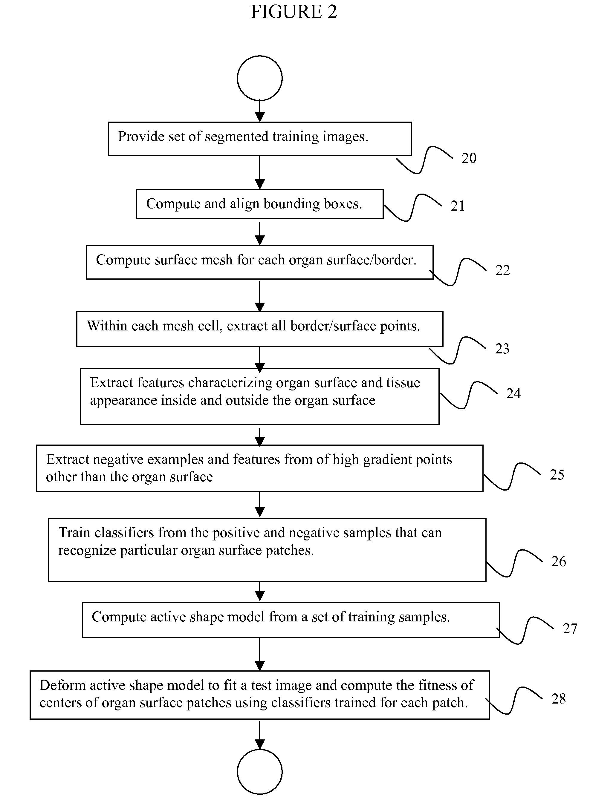
A method for segmenting organs in digitized medical images includes providing a set of segmented training images of an organ, computing a surface mesh having a plurality of mesh cells that approximates a border of the organ, extracting positive examples of all mesh cells and negative examples in the neighborhood of each mesh cell which do not belong to the organ surface, training from the positive examples and negative examples a plurality of classifiers for outputting a probability of a point being a center of a particular mesh cell, computing an active shape model using a subset of center points in the mesh cells, generating a new shape by iteratively deforming the active shape model to fit a test image, and using the classifiers to calculate a probability of each center point of the new shape being a center of a mesh cell which the classifier was trained to recognize.

Description

CROSS REFERENCE TO RELATED UNITED STATES APPLICATIONS[0001]This application claims priority from “Organ segmentation using surface patch classification in 2D and 3D images”, U.S. Provisional Application No. 60 / 977,120 of Jerebko, et al., filed Oct. 3, 2007, the contents of which are herein incorporated by reference in their entirety.TECHNICAL FIELD[0002]This disclosure is directed to techniques for organ segmentation in digital medical images, including, but not limited to, images of computed tomography (CT), magnetic resonance imaging (MRI), positron emission tomography (PET), and other modalities.DISCUSSION OF THE RELATED ART[0003]The diagnostically superior information available from data acquired from current imaging systems enables the detection of potential problems at earlier and more treatable stages. Given the vast quantity of detailed data acquirable from imaging systems, various algorithms must be developed to efficiently and accurately process image data. With the aid of...

Claims


Application Information

Patent Timeline
IPC IPC(8): G06K9/00
CPCG06T7/0083G06T7/0089G06T17/20G06T2207/30004G06T2207/20081G06T2207/20124G06T2207/10072G06T7/12G06T7/149
Inventor JEREBKO, ANNAWOLF, MATTHIAS
Owner SIEMENS HEATHCARE GMBH
Why Eureka
Try Eureka