Method and system for dynamic pulmonary trunk modeling in computed tomography and magnetic resonance imaging

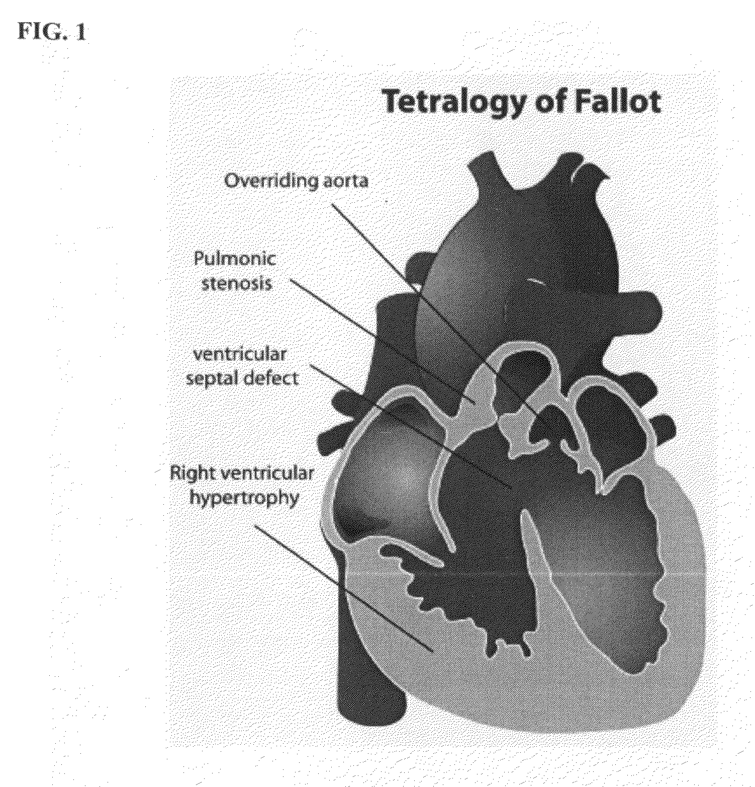
a computed tomography and magnetic resonance imaging technology, applied in image enhancement, diagnostic recording/measuring, instruments, etc., can solve the problems of pulmonary valve diseases, pulmonary insufficiency, and pulmonary valve damag

Active Publication Date: 2009-06-18
SIEMENS HEALTHCARE GMBH
View PDF10 Cites 29 Cited by
  • Summary
  • Abstract
  • Description
  • Claims
  • Application Information

AI Technical Summary

Problems solved by technology

Although left-side (e.g., aortic) valves are affected more often, pulmonary valve diseases also represent a serious health problem.
Such conditions require constant monitoring and, at some stage, typically require valve intervention.
This initial intervention can damage or in some cases destroy the pulmonary valve leading to pulmonary insufficiency.
The timing for this re-intervention procedure is not well defined and requires constant monitoring of the patient.
However, the main difficulty of PPVI is that it is difficult to assess the pulmonary trunk before treatment.
Due to this difficulty, management of patients with pulmonary valve disease has remained challenging.
This is inefficient and in many cases is not sufficiently accurate due to the complexity of the pulmonary trunk anatomy.
Manually determining the same relative axial position within the pulmonary trunk can often lead to biased measurements.
Furthermore, two dimensional projections cannot capture through plane motion.
Moreover, elaborate functional quantifications are not possible using conventional methods.

Method used


Image

  • Method and system for dynamic pulmonary trunk modeling in computed tomography and magnetic resonance imaging
  • Method and system for dynamic pulmonary trunk modeling in computed tomography and magnetic resonance imaging
  • Method and system for dynamic pulmonary trunk modeling in computed tomography and magnetic resonance imaging

Examples


Embodiment Construction

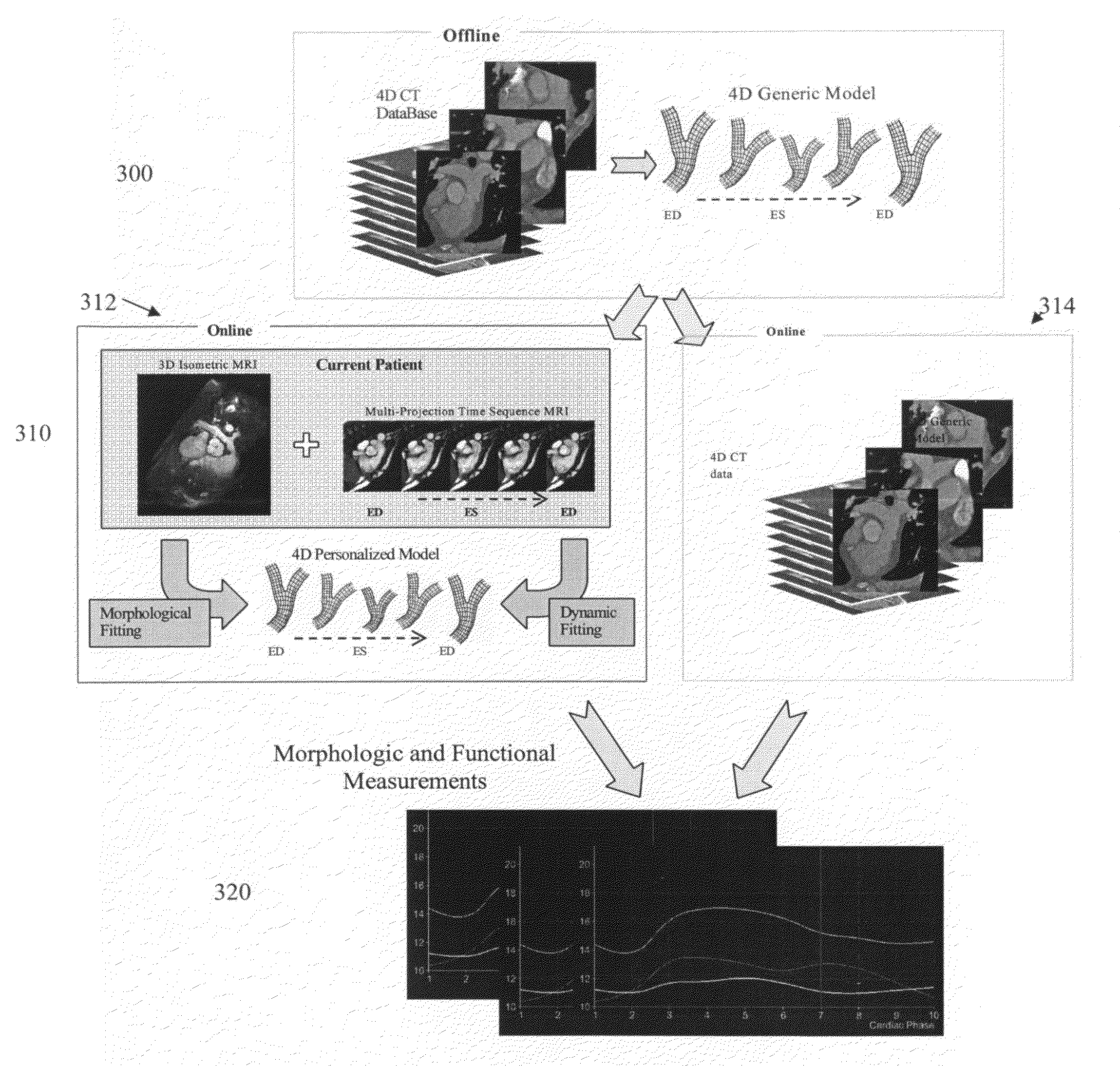
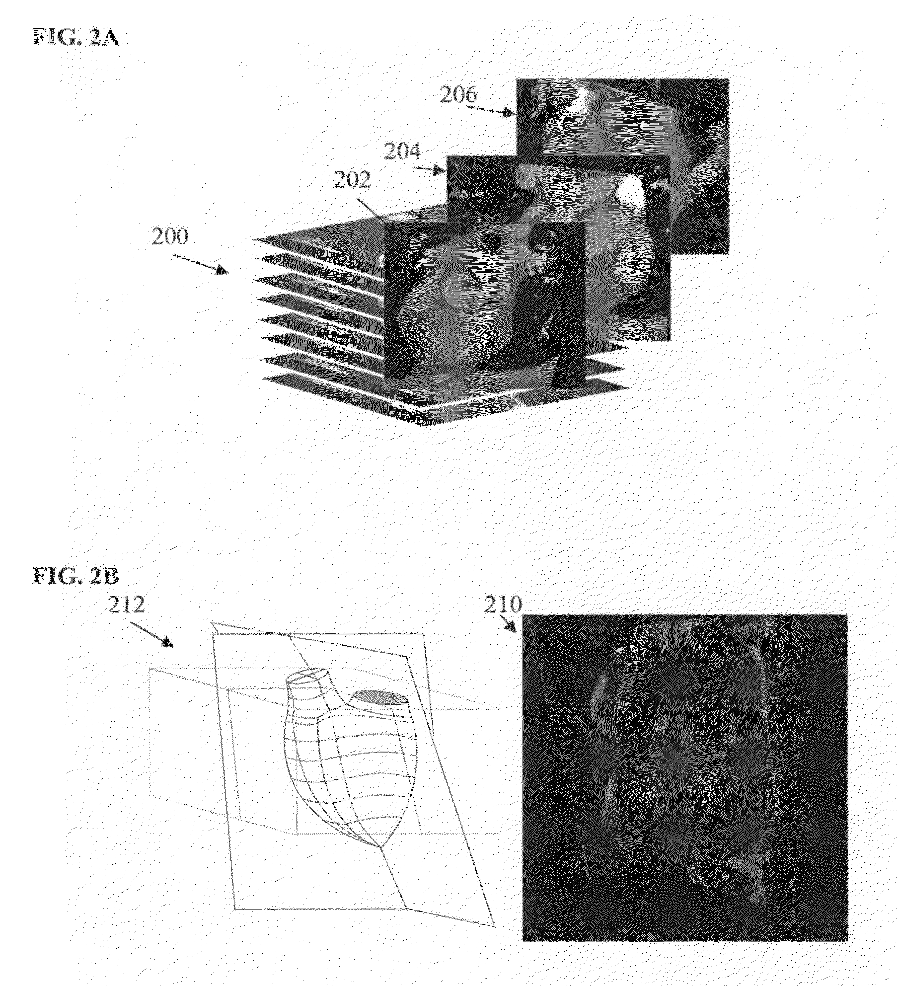
[0021]The present invention relates to modeling and quantitative evaluation of the pulmonary trunk using 4D computed tomography (CT) or magnetic resonance imaging (MRI) data. Embodiments of the present invention are described herein to give a visual understanding of the heart modeling method. A digital image is often composed of digital representations of one or more objects (or shapes). The digital representation of an object is often described herein in terms of identifying and manipulating the objects. Such manipulations are virtual manipulations accomplished in the memory or other circuitry / hardware of a computer system. Accordingly, is to be understood that embodiments of the present invention may be performed within a computer system using data stored within the computer system. Embodiments of the present invention are described herein as using 4D CT data or 4D MRI data to model and quantitatively evaluate the aortic valve. It is to be understood that the present invention is ...


PUM


Abstract

A method and system for modeling the pulmonary trunk in 4D image data, such as 4D CT and MRI data, is disclosed. Bounding boxes are detected in frames of the 4D image data. Anatomic landmarks are detected in the frames of the 4D image data based on the bounding boxes. Ribs or centerlines of the pulmonary artery are detected in the frames of the 4D image data based on the anatomic landmarks, and a physiological pulmonary trunk model is fit the frames of the 4D image data based on the detected ribs and anatomic landmarks. The boundary of the pulmonary trunk is detected in order to refine the boundary of the pulmonary trunk model in the frames of the 4D image data, resulting in a dynamic model of the pulmonary trunk. The pulmonary trunk can be quantitatively evaluated using the dynamic model.

Description

[0001]This application claims the benefit of U.S. Provisional Application No. 61 / 013,018, filed Dec. 12, 2007, the disclosure of which is herein incorporated by reference.BACKGROUND OF THE INVENTION[0002]The present invention relates to dynamic modeling of the pulmonary trunk using medical images, and more particularly, to modeling and quantitative evaluation of the pulmonary trunk using 4D computed tomography (CT) and magnetic resonance imaging (MRI) data.[0003]Valvular heart disease (VHD) is a cardiac disorder that affects a large number of patients and often requires elaborate diagnostic procedures, intervention, and long-term management. Although left-side (e.g., aortic) valves are affected more often, pulmonary valve diseases also represent a serious health problem. In most case, pulmonary abnormalities occur in conjunction with other heart disease and can be caused by congenital defects, pulmonary hypertension, endocarditis, rheumatic fever, and carcinoid heart disease. Such c...

Claims


Application Information

Patent Timeline
Patent Type & Authority Applications(United States)
IPC IPC(8): G06T7/00
CPCG06T7/0028G06T7/0038G06T2207/30048G06T2207/10081G06T2207/10088G06T2207/10076G06T7/33G06T7/38
Inventor LYNCH, MICHAELIONASEC, RAZVANGEORGESCU, BOGDANCOMANICIU, DORINVITANOVSKI, DIME
Owner SIEMENS HEALTHCARE GMBH
Why Eureka
Try Eureka