Method and user interface for the graphical presentation of medical data

Inactive Publication Date: 2009-10-08
SIEMENS AG
View PDF5 Cites 4 Cited by
  • Summary
  • Abstract
  • Description
  • Claims
  • Application Information

AI Technical Summary

Benefits of technology

[0018]An advantage of the invention is that detail and context information can be presented simultaneously. This reduces the cognitive effort for the user, and also reduces the time needed for evaluation and assessment since no switching between the views need occur. Such a mode of operation accommodates visually oriented people. The presentation can be adapted without further measures to various

Problems solved by technology

A simultaneous presentation of detail view and context view is not possible.
The access or the presentation of the detail view and a context view

Method used


Image

  • Method and user interface for the graphical presentation of medical data
  • Method and user interface for the graphical presentation of medical data
  • Method and user interface for the graphical presentation of medical data

Examples


Embodiment Construction

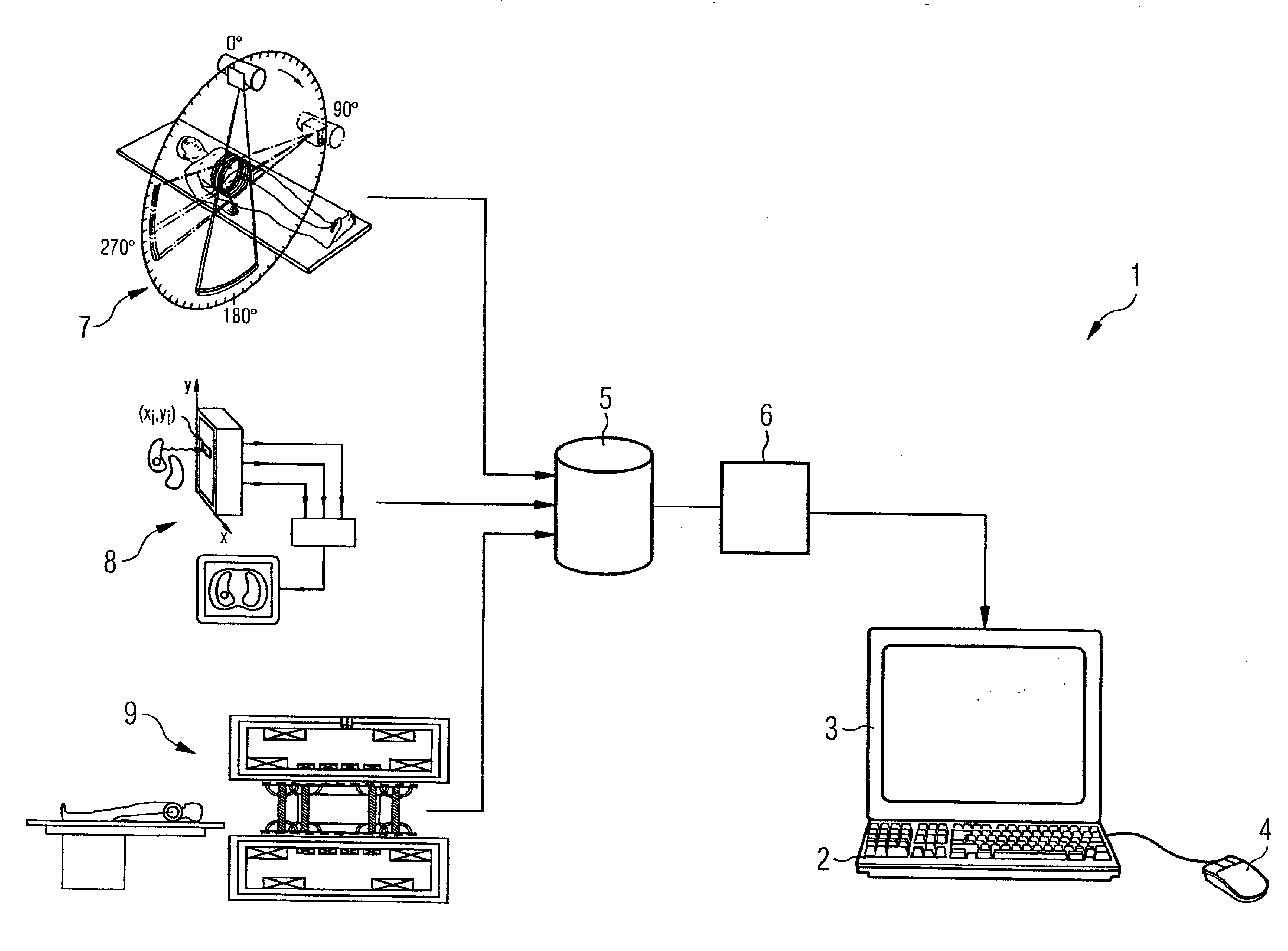
[0023]The presentation in the figures is not to scale; identical elements or elements having identical effects are provided with the same reference characters.

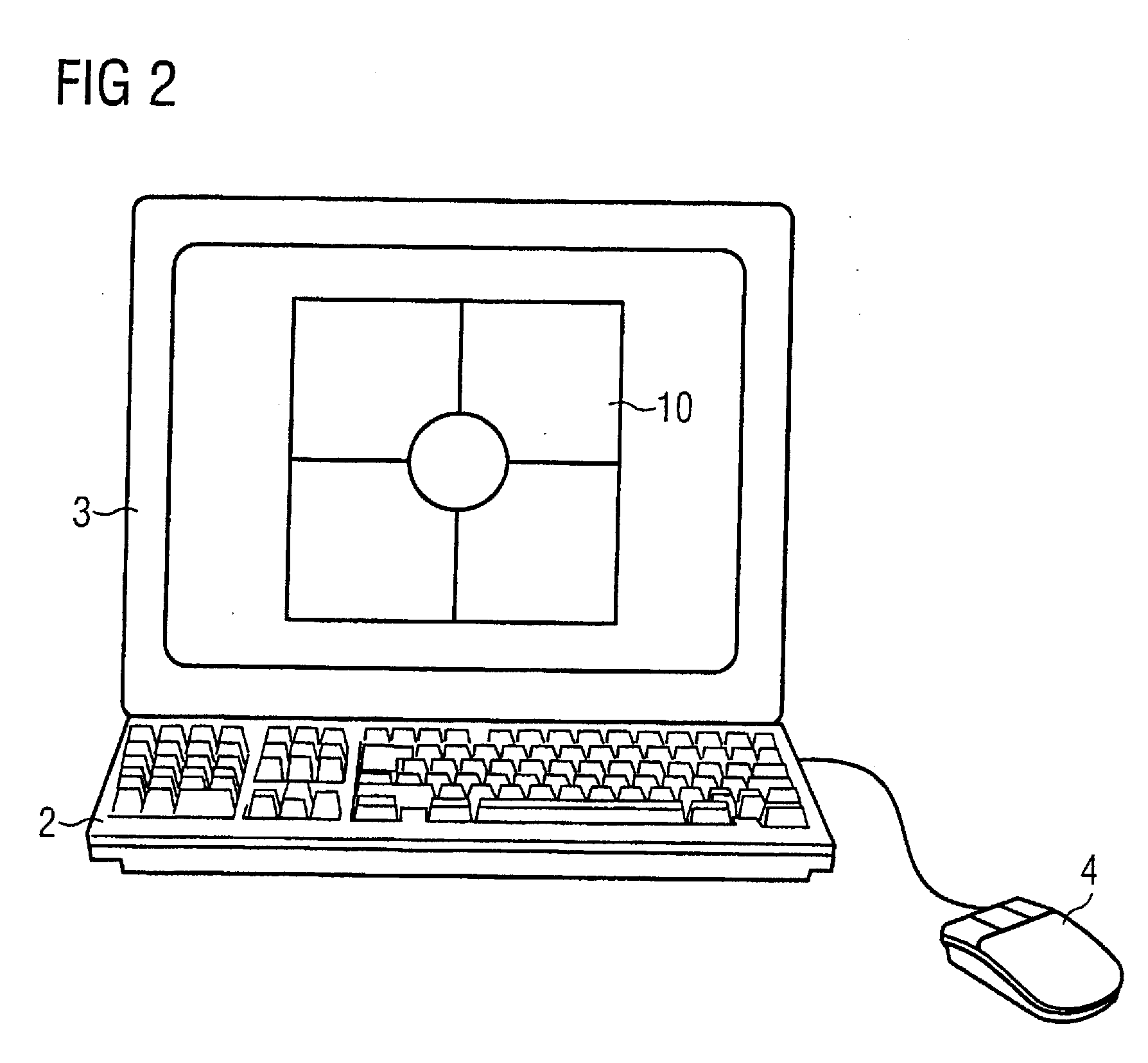
[0024]The user interface according to the invention utilizes the possibilities of the display of an additional element of the user interface (known as a “UI element”) which can be superimposed on the original view. Via this additional element of the user interface, the user now has the possibility to receive context information as usual as well as to simultaneously receive detail information of the data object in the UI element.

[0025]The region of a data object that is focused on by the user can be enlarged or, respectively, reduced in the UI element. The factor of the enlargement can be continuously adjustable. The selection of the region of the data object to be shown in the UI element can by conducted by the user with the assistance of corresponding input devices. The position of the UI element on the screen is freely defin...


PUM


Abstract

Operation of a method and a device for the implementation of a medical examination via a user interface of at least one imaging device include determination and input of a group of measurement parameters via an input device of the user interface, generation of spatially resolved image information with the at least one imaging device depending on the group of measurement parameters, and storage of the image information on a storage medium and presentation of the image information as a data symbol on a screen of the user interface. Multiple data symbols are presented in a predetermined arrangement on the screen for a medical assessment. A UI element on the screen and a pointer symbol is positioned on the screen in one of the multiple data symbols via an input device. A predetermined region at the position of the pointer symbol is shown in the UI element.

Description

BACKGROUND OF THE INVENTION[0001]1. Field of the Invention[0002]The present invention concerns the graphical presentation of medical examination results that were acquired with imaging methods, and in particular a method and a user interface for the implementation of a medical examination.[0003]2. Description of the Prior Art[0004]In the implementation of imaging methods in medical technology (for example magnetic resonance tomography, computed tomography etc.), large sets (quantities) of data (radiological data sets) are regularly acquired. Various methods (known as browsers) are known from the prior art for the necessary consideration, processing and assessment of the acquired data. These browsers for radiological data sets allow the user (radiologist, MRTA) to view the representation of the data sets in detail views or context views.[0005]The user interfaces of browsers for radiological data sets are typically designed for a specific user situation. For example, in the browsers i...

Claims


Application Information

Patent Timeline
IPC IPC(8): G06F3/048G16Z99/00
CPCA61B6/467A61B6/469A61B6/5235G06T19/00A61B19/56G06F19/3406A61B6/566A61B34/25G16H40/63G16Z99/00
Inventor GLASER-SEIDNITZER, KARLHEINZKLING, JOHANNESREQUARDT, MARTIN
Owner SIEMENS AG
Why Eureka
Try Eureka