
Method and device for image guided surgery

a technology of image guided surgery and a device, applied in the field of image guided surgery, can solve the problems of adding to overall care costs and exposing patients to potential nosocomial infections

Inactive Publication Date: 2010-02-11
WEINBERG MEDICAL PHYSICS
View PDF7 Cites 3 Cited by
  • Summary
  • Abstract
  • Description
  • Claims
  • Application Information

AI Technical Summary

Benefits of technology

[0021]It is, therefore, one aspect of the present invention to provide an apparatus that permits a physician and / or surgeon to identify the margins between healthy and unhealthy tissue during a surgical procedure.
[0024]Often, patients reside in hospital beds while pathological assessment of tissue specimens is pending, adding to overall care costs and exposing patients to potential nosocomial infection. A rapid and reliable intraoperative method of assessing surgical margins could potentially reduce hospital stays. Moreover, providing additional confidence might allow surgeons and podiatrists to be more conservative in their resections, thereby improving patients' quality of life.
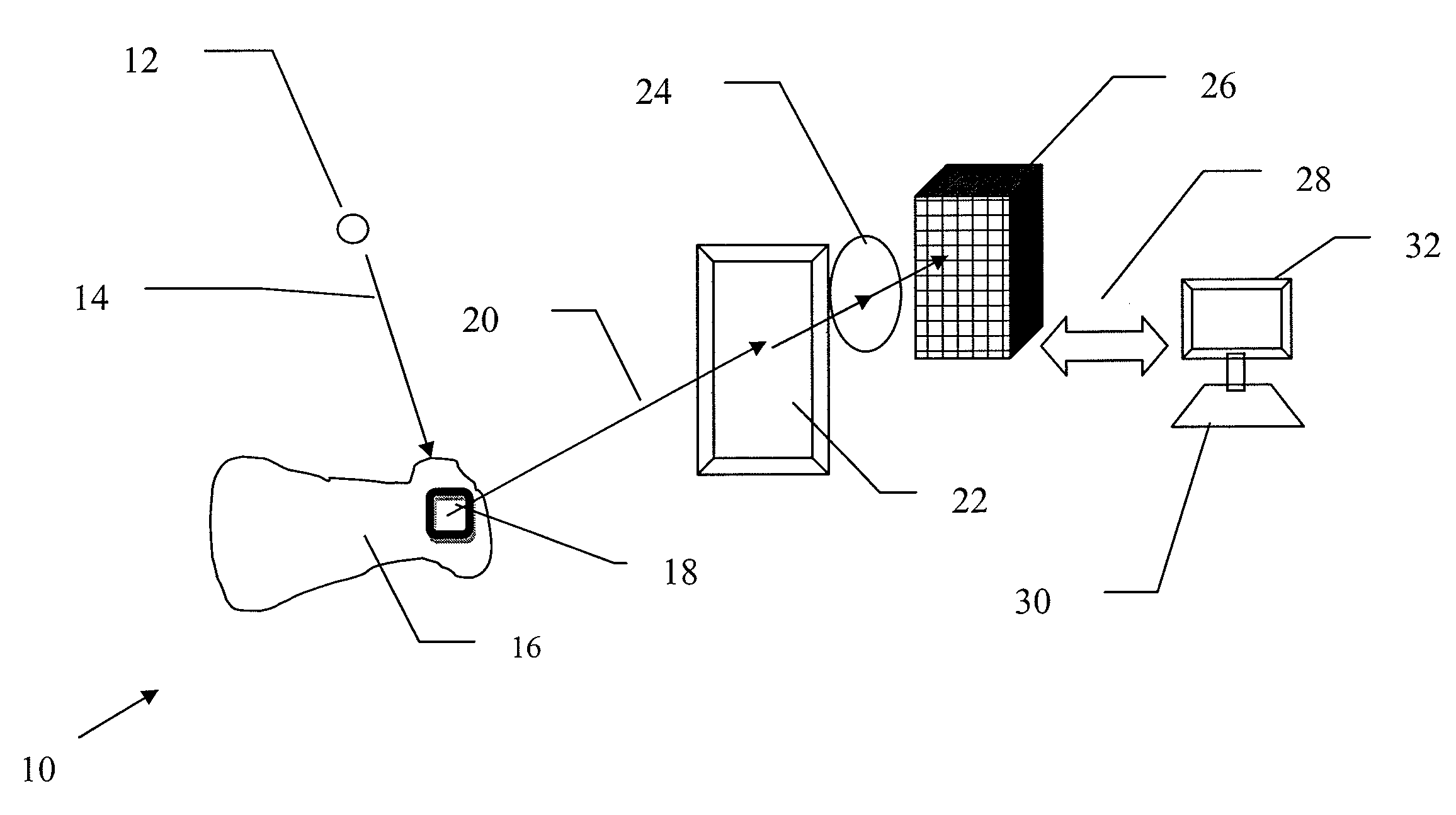
[0025]Unlike prior imaging approaches that employed exogenous contrast materials (e.g., tetracycline, labeled radiotracers) the present invention exploits the availability of endogenous markers (e.g., NADH, which is the reduced form of nicotinamide adenine dinucleotide (“NAD”)) to rapidly assess tissue viability.

Problems solved by technology

Often, patients reside in hospital beds while pathological assessment of tissue specimens is pending, adding to overall care costs and exposing patients to potential nosocomial infection.

Method used


Image

  • Method and device for image guided surgery
  • Method and device for image guided surgery
  • Method and device for image guided surgery

Examples


Embodiment Construction

[0041]The present invention will now be described in connection with one or more embodiments. The discussion of specific embodiments, however, is not intended to be limiting of the invention. To the contrary, the selected embodiments are intended to be exemplary of the broad scope of the present invention. As should be appreciated by those skilled in the art, there are numerous variations and equivalents that may be employed without departing from the present invention. Those embodiments and variations are intended to be encompassed by the present invention.

[0042]While the present invention is described primarily in connection with the identification of healthy and unhealthy tissue in the context of a diabetic patient, it is contemplated that the present invention will be applicable to a wide variety of circumstances.

[0043]As indicated above, fluorescence is exhibited naturally by many organisms, primarily from NADH. NADH is component of the Krebs cycle, with broad application to bi...


PUM


Abstract

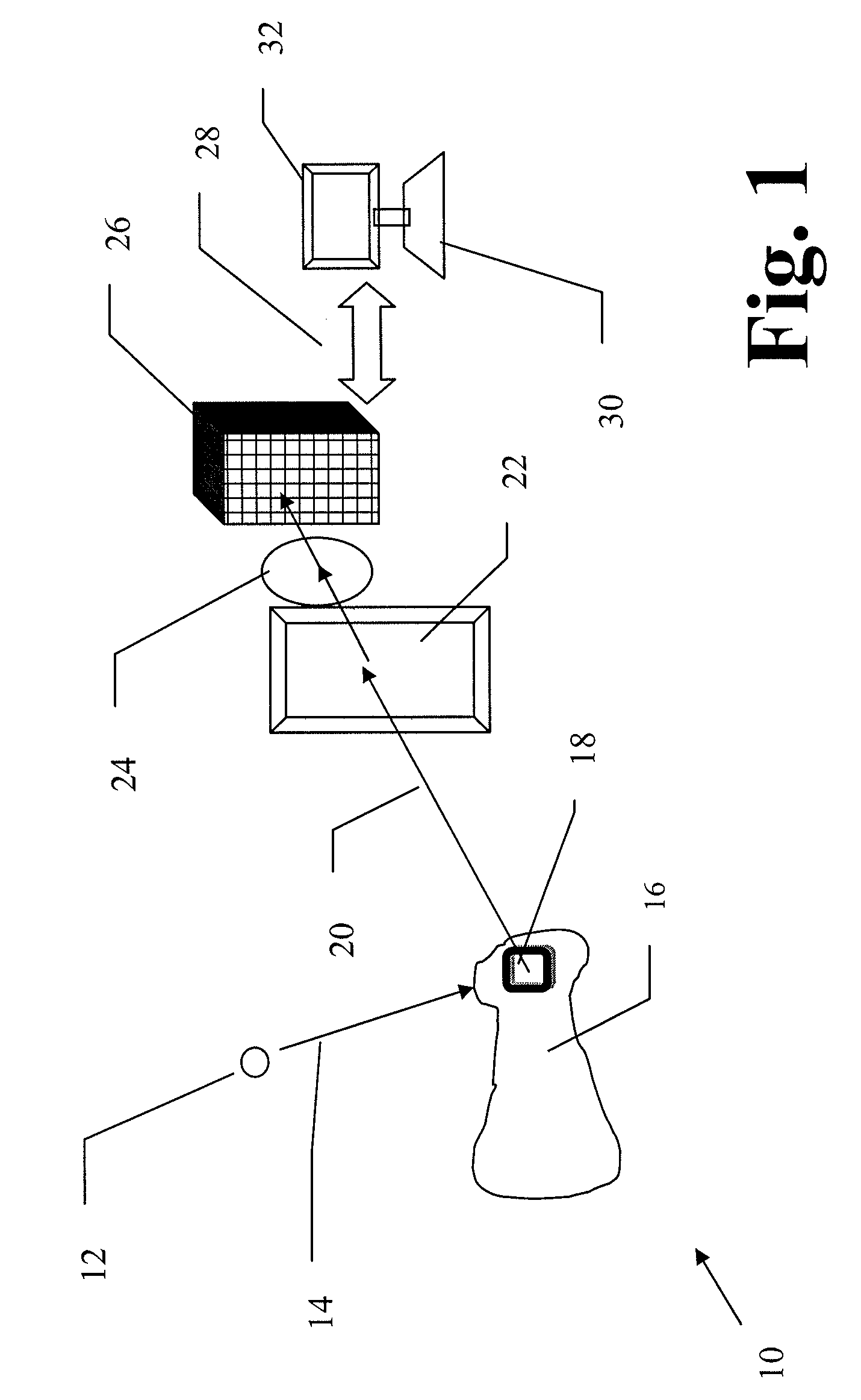
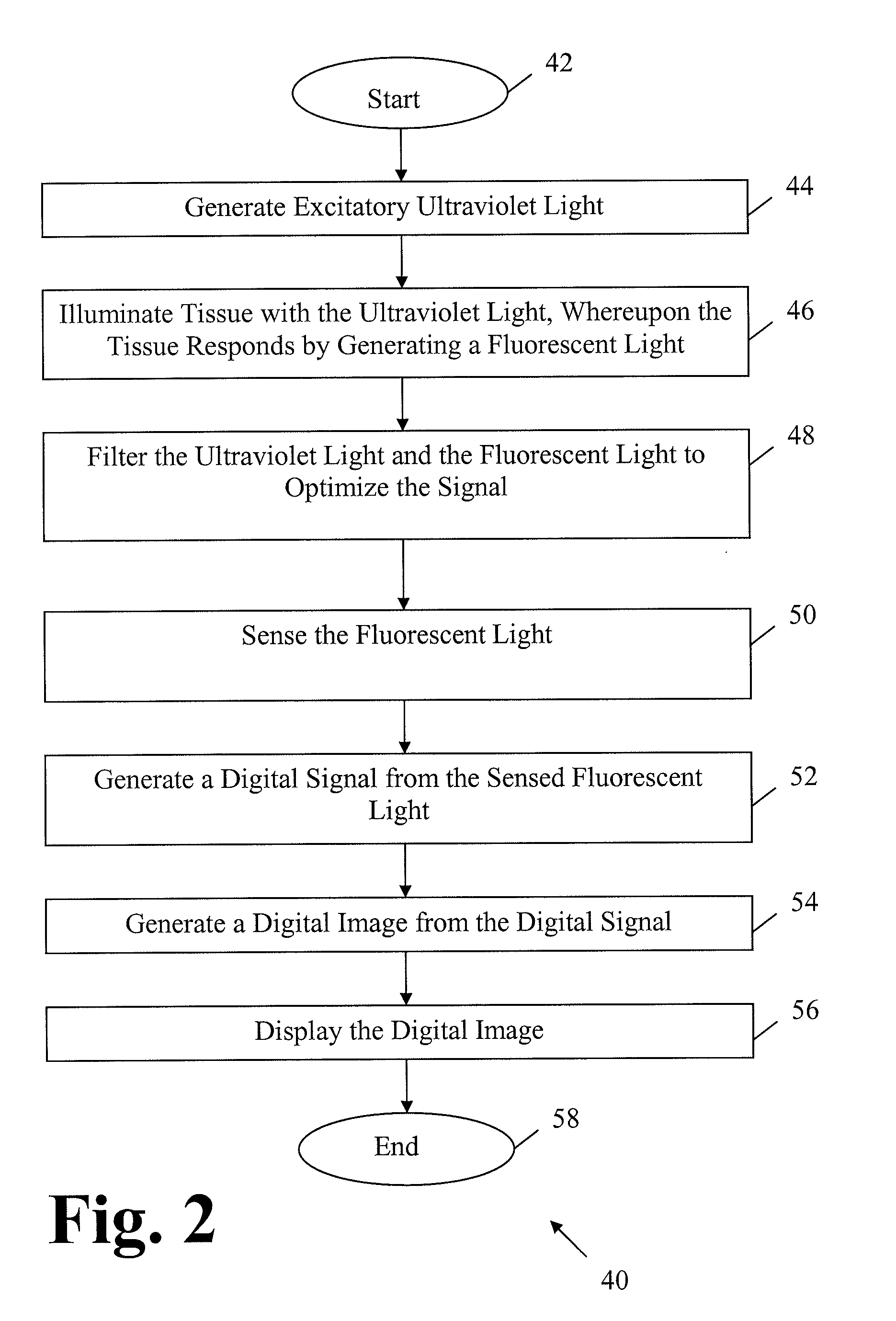
A fluorometer includes a light source to generate excitatory light toward a tissue, the tissue generating fluorescent light in response to the excitatory light. The fluorometer also includes a light sensor to receive the fluorescent light and generate a digital signal. A processor is connected to the light sensor to receive the digital signal and generate a digital image, and a display displays the digital image. The tissue generates fluorescent light as a result of excitation of at least one intrinsic tissue metabolic product. A method for distinguishing between viable and non-viable tissue using the fluorometer also is described.

Description

CROSS-REFERENCE TO RELATED APPLICATION(S)[0001]This is a United States Non-Provisional patent application that relies for priority on U.S. Provisional Patent Application No. 61 / 086,402, filed on Aug. 5, 2008, the contents of which are incorporated herein by reference.BACKGROUND OF THE INVENTION[0002]The present invention involves an apparatus and a method that provides intraoperative imaging feedback to permit a podiatrist and / or surgeon to determine the margins between healthy and unhealthy tissue. More specifically, the present invention provides an apparatus and a method that permits imaging of tissue so that a surgeon may determine the margins between healthy and unhealthy tissue, especially in circumstances where the unhealthy tissue is to be removed surgically.DESCRIPTION OF THE RELATED ART[0003]Generally speaking, diabetic patients are prone to a number of maladies associated with diabetes. Since diabetes results in a generally elevated glucose concentration in the patients b...

Claims


Application Information

Patent Timeline
Patent Type & Authority Applications(United States)
IPC IPC(8): A61B6/00
CPCA61B5/0059A61B2019/5231A61B19/5225A61B5/411A61B90/37A61B2090/373
Inventor WEINBERG, IRVINGSTEPANOV, PAVEL
Owner WEINBERG MEDICAL PHYSICS
Learn More