Head-stabilized medical apparatus, system and methodology

a technology of stabilizing medical equipment and head, applied in the field of head stabilizing medical equipment, can solve the problems of inability to achieve optimal effectiveness and accuracy of eng/vng test batteries, unable to employ ideal anatomical positions to obtain useful information, and unable to achieve optimal effectiveness and accuracy.

Inactive Publication Date: 2010-02-18
VESTICON BSI
View PDF43 Cites 16 Cited by
  • Summary
  • Abstract
  • Description
  • Claims
  • Application Information

AI Technical Summary

Benefits of technology

[0004] In a manner of speaking, the invention recognizes, and centers attention on, the discovered significance of utilizing various, plural-simultaneously-employed sensors / detectors which are specially positionally stabilized, both (a) with respect to the head of a patient, and (b) with respect to each other, for the simultaneous gathering, and immediate computer processing, of plural-parameter data which can lead to accurate diagnoses and treatment of disorders of the types just generally mentioned above. Both mentioned categories of stabilization have been found to be important and unique in this sophisticated and challenging field of medical practice. Positional stabilization, undertaken in accordance with practice of the invention, leads to accurate correlation of different simultaneously gathered data components, and thus leads, in turn, to significant improvements in diagnostic speed and accuracy, and in trustable opportunities to rely with confidence on rapid, computer-based vestibular analyses and conclusions.
[0009] What is needed, and is definitively provided, among other things, by the present invention, is a method of carrying out the indicated screening and selected tests that can be automated and programmed to carry out certain screening tests and, but that is (a) capable of interjecting certain more definitive tests when so indicated by the screening tests, (b) can perform tests that are physiologically more meaningful than those previously done, and useful in diagnosing and treating a subject, (c) can acquire real-time data in a continuum throughout a test session, (d) can distinguish between normal and abnormal nystagmus, and (e) can, through careful programming, accomplish these tasks in as short a time as possible. Means must also be available that selects, analyzes and displays acquired data in a brief, understandable, and reliable summary.
[0016] One example of this is seen with vestibular lithiasis, or benign paroxysmal positional vertigo and different variants, whereby abnormal particles in the semicircular canals render the canals sensitive to linear acceleration, including gravitation, creating symptoms of vertigo in response to position change of the head relative to gravity. These conditions are very common, and can often be improved or corrected by repositioning maneuvers, whereby the particles are moved, via a particular sequential positioning of the subject's head with optional induced head oscillation, to an area of the labyrinth where they no longer produce abnormal responses. Most subjects with these conditions can be treated successfully by canalith repositioning maneuvers, including variations thereof, collectively known as particle repositioning maneuvers, which are designed to cause migration of aberrant particles to an area of the labyrinth where they no longer affect the dynamics of the semicircular canal.
[0019] One related and very important novel contribution of the present invention is its demonstrable ability, on-the fly, so to speak, to distinguish even very subtly existent pathological (abnormal) from physiological (normal) nystagmus events. This is extremely valuable to the clinician during testing or treatment, because of the fact that the nystagmus being observed in response to head maneuvers often contains components of both pathological and physiologic nystagmus. It is clearly advantageous to be able to observe and analyze just the pathological nystagmus without contamination by physiological nystagmus. Positional stability of sensors and stimulators in accordance with practice of the present invention leads significantly to the reliable ability to accomplish this differentiation.
[0020] Physiological nystagmus is mainly induced by angular acceleration of the head, with the slow phase of nystagmatic eye rotation occurring in the same plane as, but in the opposite direction from, head movement. This is a normal response reflex. Thus, by monitoring angular acceleration in addition to linear acceleration, while also monitoring, simultaneously, eye movement, and by doing all of this under conditions wherein the relevant monitoring sensors are firmly positionally stabilized relative both to one another, and to a patient's head, the present invention can effectively distinguish between those components of nystagmus that are physiological and those which are pathological in origin.

Problems solved by technology

Dysfunction of the semicircular canals results mainly in symptoms of vertigo.
However, these standardized test positions do not employ the ideal anatomical positions for obtaining useful information.
Inasmuch as nystagmus occurring in a particular test position will be dependent upon numerous factors, such as (a) the rapidity and method of the just-mentioned maneuver, (b) the time lapse after a test position is reached until the data-acquisition run is commenced, and (c) the exact angles of the test positions, etc, the usual ENG / VNG test battery, as now generally carried out, is not optimally effective and accurate.
Abnormal conditions adversely affecting the otolithic organs cause mainly symptoms and signs of imbalance.
This imbalance of otolithic origin results from either unstable neural input from an otolithic organ, or organs, or a bilateral deficit.
Central compensation generally takes place adequately over time for the reduced form if it is unilateral and becomes stable, but compensation is delayed or not forthcoming in response to the distorted form because of its persistent instability.
A problem with analyzing adverse postural effects for this purpose is that test subjects are usually acutely aware of their recent postural misperceptions that have resulted in abnormal sway or fall in a particular direction, and can quickly compensate for these misperceptions to some degree when presented with the same apparent stimulus.
This tendency to compensate results in limited repeatability, and thus, questionable reliability of such a test using postural control as a measure.
Aberrant receptivity of the labyrinth to sound or intralabyrinthine pressure change can occur in either the semicircular canals, thereby adversely impacting the VOR system and producing nystagmus, or in the otolithic organs, thereby adversely impacting the VSR system and producing abnormal postural effects and altered gravitational perception.
In the present state-of-the-art, quantitative information on the status of both the VOR and the VSR requires two separate devices, taking up more space in the vestibular laboratory and adding to expense.
In addition, several valuable existing tests have not been utilized significantly outside of research laboratories because of the expense involved in the equipment to perform each test separately.
However, for the more complicated cases, optimal performance of these maneuvers requires ongoing, real time observation and analysis of nystagmus.
In addition, and as mentioned above, the nystagmus patterns that subjects may display in response to maneuvers may be rapidly changing and complex, yet immediate interpretation is often required, and this requirement becomes more acute when the need for a change in strategy is indicated (e.g. a conversion of the causative particles from the posterior to the horizontal canal, or the development of a jamming of the particles).
Very challenging, there is the need for the operator, during an entire sequence of maneuvers, to envision the 3-D orientation, with respect to space and gravity, of the semicircular canals inside the head, as well as the apparent position of the particles within those canals.
This multi-level observation requirement is quite difficult because of the constant changing orientation in space of the subject during maneuvers.

Method used


Image

  • Head-stabilized medical apparatus, system and methodology
  • Head-stabilized medical apparatus, system and methodology
  • Head-stabilized medical apparatus, system and methodology

Examples


Embodiment Construction

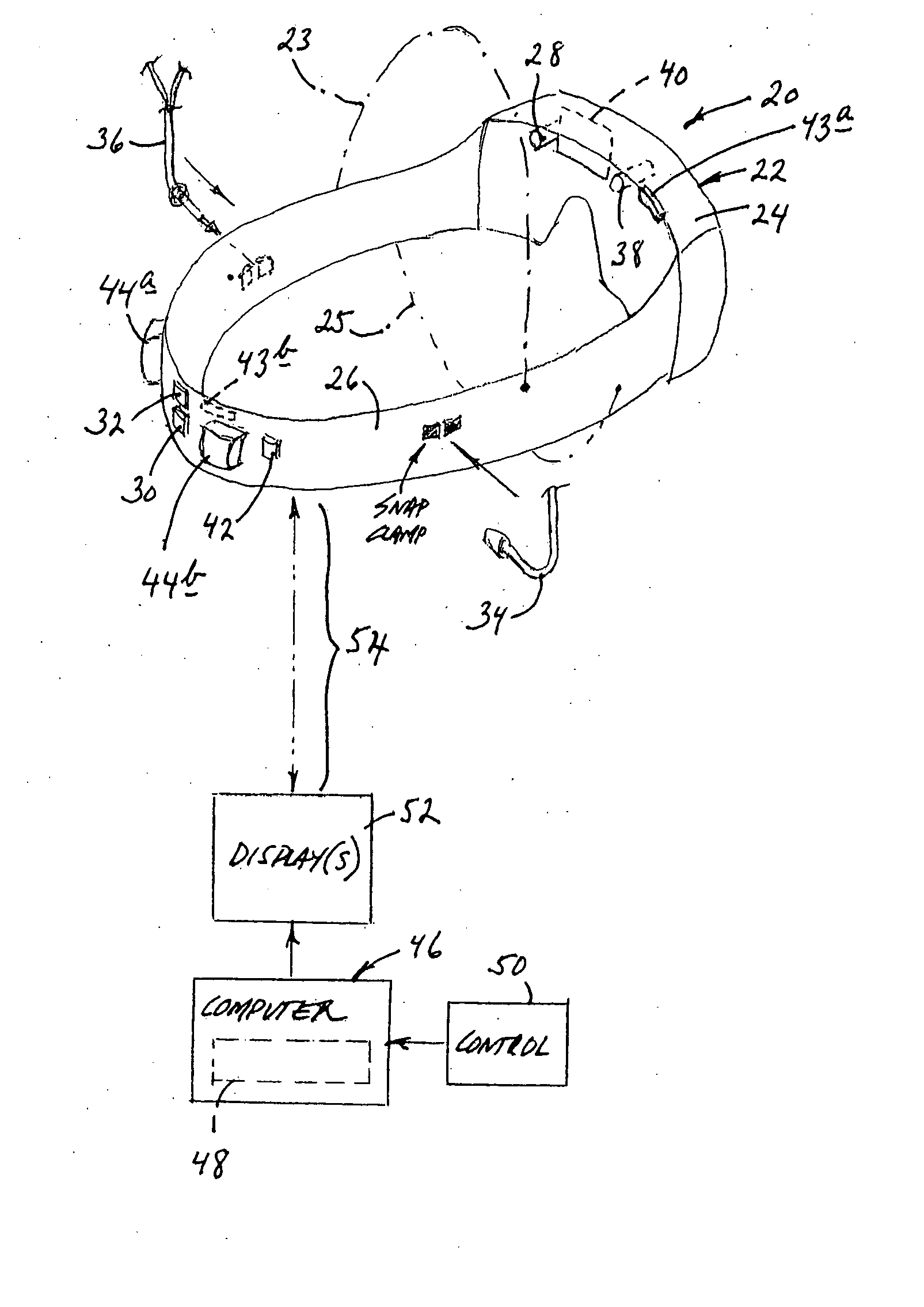
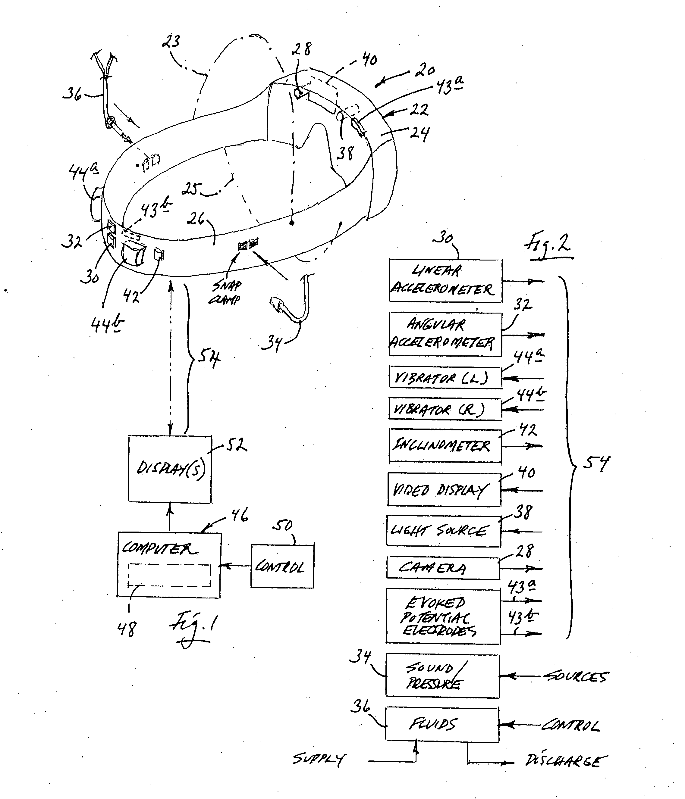
[0040] As has been mentioned above, the present invention, from a structural point of view, takes the form generally of apparatus for assisting in the computer-aided, substantially real-time diagnoses and treatments of vestibular disorders. That apparatus features head-wearable frame structure that is adapted for wearing on a subject's head in a condition of relative positional stability. The invention further features, in association with that frame structure, at least a pair of what are referred to as vestibular-parameter, data-parameter devices that are selectively anchorable to the frame structure in conditions of relative positional stability, both with respect to the frame structure, and with respect to each other. Each of these devices, in accordance with the invention, is adapted to engage in at least one of the activities which include (a) delivering to, and (b) receiving from, a subject's head vestibular-relevant parameter data. Appropriate communication structure connects...


PUM


Abstract

Structure and methodology involving mountable and head-wearable frame structure which is positionally stabilized, during use, relative a human subject's head, and which carries a selection of positionally anchored data sensors, and stimuli deliverers, that are relevant to the diagnosis and treatment of vestibular disorders. Special configurations are provided for two types of stimulators, one for sound application and air-pressure modification, and the other for the introduction of fluids to the ear. Stabilization enables tight and accurate correlation of data which is quickly analyzable by a connected, properly algorithmed computer, which can also be used for feedback control in a designed “expert” system. The invention enables, among other things, practical and significant differentiation between physiological and pathological nystagmus.

Description

[0001] This application is a Divisional of U.S. patent application Ser. No. 10 / 715,871, entitled HEAD-STABILIZED MEDICAL APPARATUS, SYSTEM AND METHODOLOGY, filed 17 Nov. 2003, the disclosure of which is herein incorporated by reference in its entirety.[0002] This invention was made with government support under PHS 398 / 2590 awarded by the National Institutes of Health. The government has certain rights in the invention.BACKGROUND AND SUMMARY OF THE INVENTION [0003] The present invention involves a head-stabilized method and apparatus designed for the diagnosis and treatment of vestibular disorders involving symptoms of dizziness, vertigo and / or imbalance. It also relates to the structures of certain special devices that are particularly suited for use with this method and apparatus, and to certain procedural approaches that the structure and method of the invention make advantageous. [0004] In a manner of speaking, the invention recognizes, and centers attention on, the discovered s...

Claims


Application Information

Patent Timeline
Patent Type & Authority Applications(United States)
IPC IPC(8): A61B5/00A61B13/00A61B5/103A61B5/113
CPCA61B3/113A61B5/0484A61B5/0496A61B5/11A61B5/1116A61B5/1126A61B5/6831A61B5/4839A61B5/4863A61B5/6814A61B5/7264G06F19/3418A61B5/113A61B5/4023A61B5/377A61B5/398G16H20/30G16H40/67
Inventor EPLEY, JOHN M.
Owner VESTICON BSI
Why Eureka
Try Eureka