Computer aided diagnostic system incorporating lung segmentation and registration

a computer-aided diagnostic system and lung nodule technology, applied in image analysis, image enhancement, instruments, etc., can solve the problems of difficult to distinguish true nodules from shadows, vessels and ribs, and difficult to detect pulmonary nodules with computer-aided image data search schemes, etc., to achieve the effect of changing the volume of a nodul

Active Publication Date: 2010-05-06
UNIV OF LOUISVILLE RES FOUND INC
View PDF6 Cites 74 Cited by
  • Summary
  • Abstract
  • Description
  • Claims
  • Application Information

AI Technical Summary

Benefits of technology

[0017]The invention addresses these and other problems associated with the prior art by providing a computer aided diagnostic system and automated method for diagnosing lung cancer through tracking of the growth rate of detected pulmonary nodules over time. Embodiments consistent with the invention determine the growth rate between first and second chest scans taken at first and second times by segmenting out a nodule from its surrounding lung tissue and calculating the volume of the nodule only after the image data for lung tissue (which also includes image data for a nodule) has been segmented from the chest scans and the segmented lung tissue from the chest scans has been globally and locally aligned to compensate for positional variations in the chest scans as well as variations due to heartbeat and respiration during scanning. By doing so, the change in volume of a nodule between the two chest scans can be more accurately obtained.

Problems solved by technology

Early detection of lung tumors (visible on chest film as pulmonary nodules) may increase the patient's chance of survival, but detecting pulmonary nodules is a complicated task.
The difficulty for computer aided image data search schemes is distinguishing true nodules from (overlapping) shadows, vessels and ribs.
Regular chest radiography and sputum examination programs have proven ineffective in reducing mortality rates.
Although screening for lung cancer with chest X-rays can detect early lung cancer, such screening can also possibly produce many false-positive test results, causing needless extra tests.
Furthermore, while large (e.g., greater than 1 cm in diameter) malignant nodules are often relatively easy to detect with conventional screening equipment and can be diagnosed with needle biopsy or bronchoscopy techniques, these techniques are typically unsuitable for detecting smaller nodules, particularly if such nodules are located deep in the lung tissue or away from large airways.
Thus, many of these techniques have been found to be unsuitable for early stage lung cancer detection.
However, these spherical, cylindrical, or circular assumptions are typically not adequate for describing the general geometry of the lesions.
The drawbacks to these approaches are the difficulties in detecting lung wall nodules.
Two specific issues arising in connection with pulmonary nodule detection include image segmentation and registration.
Typical conventional techniques are based on fitting a Gaussian model to empirical data, but this approach becomes a challenge if initial measurements are corrupted with outliers and margin-truncation from neighboring structures.
Other techniques segment 2D and 3D nodules based on thresholding voxel intensity; however, such attempts may be unreliable on cavity or non-solid nodules.
Tracking growth from image data, however, can be a challenging task not only because of changes in a patient's position at each data acquisition, but also because of a patient's heartbeat and respiration during data acquisition.
Conventional approaches, however, have been found to inadequately account for large deformations of lung tissues due to heartbeat and respiration.
Furthermore, these approaches have been found to not be suitable for some types of pulmonary nodules such as cavities and ground glass nodules, and many approaches require significant user interaction, which can be problematic in clinical settings.

Method used


Image

  • Computer aided diagnostic system incorporating lung segmentation and registration
  • Computer aided diagnostic system incorporating lung segmentation and registration
  • Computer aided diagnostic system incorporating lung segmentation and registration

Examples


working example

Lung Registration Working Example

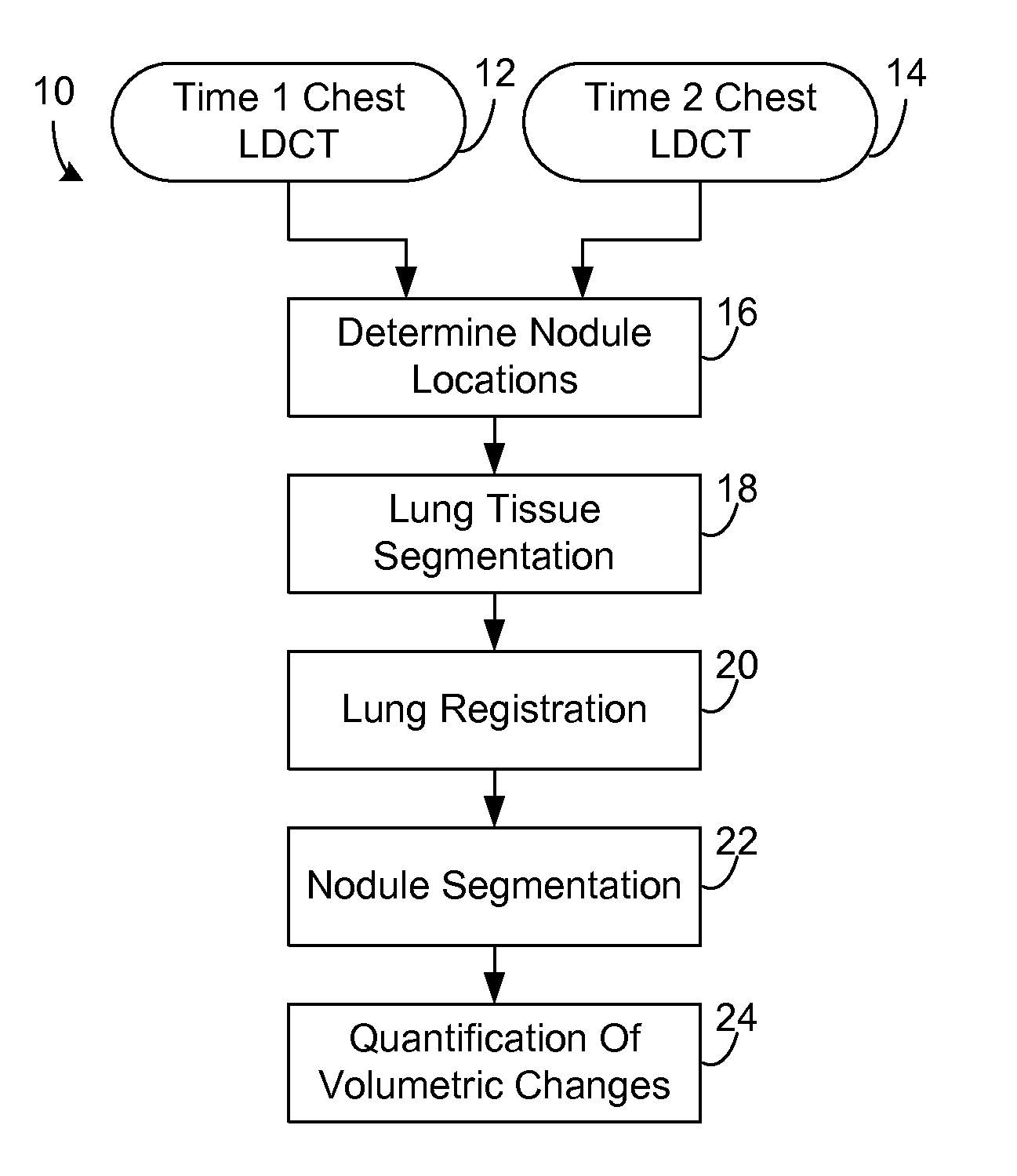
[0139]The disclosed registration techniques were tested on clinical data sets collected from 27 patients. Each patient had five LDCT scans, with a 3-month period between each two successive scans. This preliminary clinical database was collected by the LDCT scan protocol using a multidetector GE Light Speed Plus scanner (General Electric, Milwaukee, USA) with the following scanning parameters: slice thickness of 2.5 mm reconstructed every 1.5 mm; scanning pitch 1.5 mm; 140 KV; 100MA; and a field-of-view of 36 cm. After the two volumes at different time instants were registered, the lung nodules were segmented after registration using A. Farag, A. El-Baz, G. Gimel'farb, R. Falk, M. Abou El-Ghar, T. Eldiasty, S. Elshazly, “Appearance models for robust segmentation of pulmonary nodules in 3D LDCT chest images,” Proceedings of the International Conference on Medical Image Computing and Computer-Assisted Intervention (MICCAI'06), vol. 1, Copenhagen, Denma...


PUM


Abstract

A computer aided diagnostic system and automated method diagnose lung cancer through tracking of the growth rate of detected pulmonary nodules over time. The growth rate between first and second chest scans taken at first and second times is determined by segmenting out a nodule from its surrounding lung tissue and calculating the volume of the nodule only after the image data for lung tissue (which also includes image data for a nodule) has been segmented from the chest scans and the segmented lung tissue from the chest scans has been globally and locally aligned to compensate for positional variations in the chest scans as well as variations due to heartbeat and respiration during scanning. Segmentation may be performed using a segmentation technique that utilizes both intensity (color or grayscale) and spatial information, while registration may be performed using a registration technique that registers lung tissue represented in first and second data sets using both a global registration and a local registration to account for changes in a patient's orientation due in part to positional variances and variances due to heartbeat and/or respiration.

Description

CROSS-REFERENCE TO RELATED APPLICATIONS[0001]This invention claims the benefit of priority of U.S. Provisional Application Ser. No. 61 / 111,460, filed on Nov. 5, 2008, which is incorporated by reference herein in its entirety.FIELD OF THE INVENTION[0002]The invention is generally related to computer analysis of medical image data, and in particular to the segmentation and registration of image data, e.g., low dose computed tomography (LDCT) chest scans.BACKGROUND OF THE INVENTION[0003]Lung cancer remains the leading cause of mortality cancer. In 1999, there were approximately 170,000 new cases of lung cancer in the U.S., where approximately one in every eighteen women and approximately one in every twelve men develop lung cancer. Early detection of lung tumors (visible on chest film as pulmonary nodules) may increase the patient's chance of survival, but detecting pulmonary nodules is a complicated task. Nodules show up as relatively low-contrast white circular objects within the lun...

Claims


Application Information

Patent Timeline
Patent Type & Authority Applications(United States)
IPC IPC(8): G06K9/00
CPCG06T7/0016G06T2207/10081G06T2207/20016G06T2207/30064G06T7/33G06T7/35G06T7/11G06T7/143
Inventor EL-BAZ, AYMAN S.
Owner UNIV OF LOUISVILLE RES FOUND INC
Why Eureka
Try Eureka