Digital Radiography Sensors

a technology of radiography and sensors, applied in the field of digital radiography (xray) sensors, to achieve the effects of reducing the refractive error in the image received, and facilitating the adjustment of the position

Active Publication Date: 2010-10-07
KHOURI LOUIE
View PDF15 Cites 4 Cited by
  • Summary
  • Abstract
  • Description
  • Claims
  • Application Information

AI Technical Summary

Benefits of technology

[0008]The present invention provides an intraoral sensor which is rotatable for proper positioning within a dental patient's oral cavity without significant interference from the cable which is connected to a computer or module containing intermediate processing circuitry. As a result, the intraoral sensor is configured to fit comfortably and in closer proximity to a target area in an intraoral cavity than currently known static sensor and cable corrections. By allowing for rotation of the sensor, not only does the sensor fit more comfortab

Problems solved by technology

Thus, unlike presently known filmless X-ray sensors, if either the

Method used


Image

  • Digital Radiography Sensors
  • Digital Radiography Sensors
  • Digital Radiography Sensors

Examples


Embodiment Construction

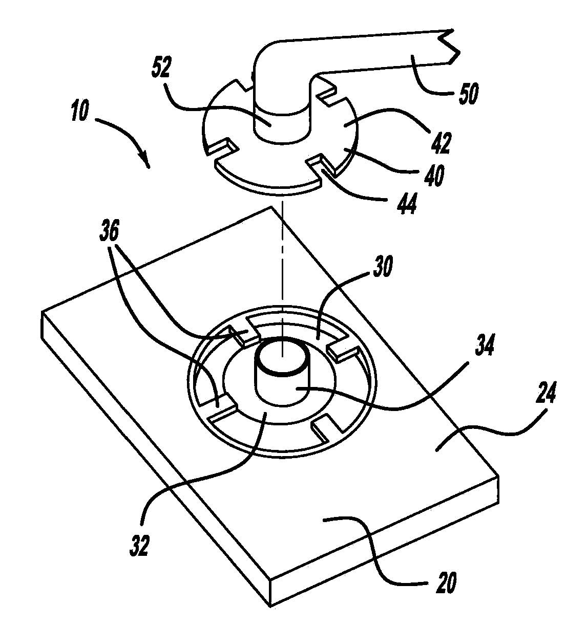
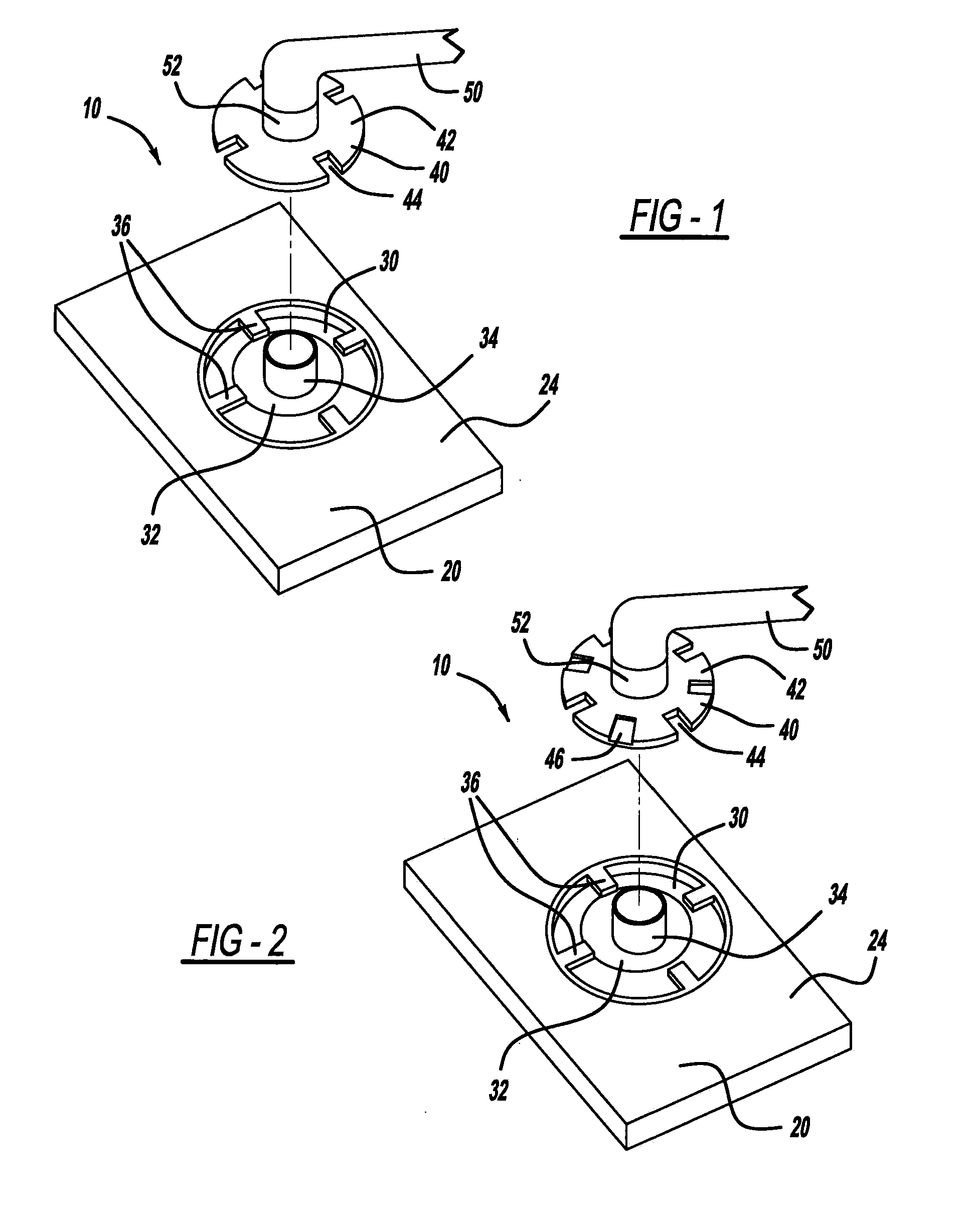
[0023]A sensor according to the present invention is designed to be used with a filmless radiography system. As an example, a sensor 10 according to the present invention can be used as part of a filmless radiography system 12 which is designed according to the principles of Schwartz U.S. Pat. No. 4,160,997 (Schwartz patent), which is incorporated hereby by reference. As illustrated in FIG. 5, the sensor 10 transmits digital image data to a preprocessor 14, and the preprocessor transmits the image data either directly to a display device 16, or to a computer 18 which is connected to the display device 16. The preprocessor 14 is configured to normalize the image data transmitted by the sensor 10, to improve the contrast (color and / or grayscale) of such data in relation to the raw image data produced by the sensor. The image data is then transmitted directly to the display device 16, or the image data is transmitted to the computer 18 which can manipulate the image data on the display...


PUM


Abstract

A new and improved digital sensor assembly for use in a filmless radiography system is disclosed. The sensor is configured to be universal in that it can be rotated about a cable connection thereby allowing the sensor to be used both vertically and horizontally in the oral cavity. The rotatability of the sensor allows it to fit comfortably and close to a target area in an intraoral cavity. By providing a comfortable relative fit to the target area, the sensor is ergonomically improved, in terms of its comfort and feel to a dental patient. Alternative sensor assembly embodiments disclosed herein relate to quick disconnect-type coupling arrangements between the sensor body and the sensor cable.

Description

BACKGROUND OF THE INVENTION[0001]The present invention relates to digital radiography (X-ray) sensors designed for use in a filmless radiography system and, more particularly, to rotatable and / or detachable digital X-ray sensors.[0002]While the present invention is considered to be applicable to both the medical and dental professions, for example, for ease in description, the following discussion will focus on an intraoral embodiment of the present invention.[0003]Dentists and oral surgeons typically use x-radiation (“X-rays”) to obtain images of their patients' teeth, mouths and gums to aid in diagnosis and treatment. In traditional oral and dental radiography, a cartridge containing a piece of photographic film is placed in the patient's mouth, for example, behind a patient's tooth, and an X-ray beam is projected through the tooth and onto the film. The film, after being exposed in this manner, is developed in a dark room or a closed processor using special chemicals to obtain a ...

Claims


Application Information

Patent Timeline
IPC IPC(8): A61B6/14
CPCA61B6/4233A61B6/145
Inventor KHOURI, LOUIE
Owner KHOURI LOUIE
Why Eureka
Try Eureka